


Contents
Calcaneal enthesophyte: symptoms and treatments
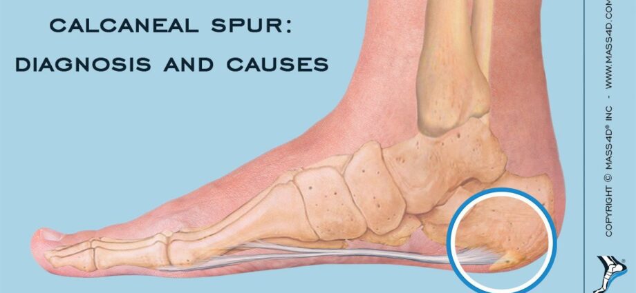
Also called calcaneal or Lenoir’s spine, the calcaneal enthesophyte is a bone growth located at the posterior part of the calcaneum, a bone located at the heel of the feet. It is caused by chronic inflammation of the plantar fascia which connects the heel to the toes and supports the entire foot. Explanations.
What is a calcaneal enthesophyte?
Thickening of the plantar fascia (a fibrous membrane that lines the entire arch of the foot), the calcaneal enthesophyte occurs in the form of a bone spine located at the posterior end of the calcaneus. It is the bone of the posterior part of the foot that constitutes the heel.
This bone spine is formed at the level of a chronic inflammation of this plantar aponeurosis, following repetitive microtraumas such as during the practice of sports which put repeated loads on the heel such as jogging, wearing shoes poorly adapted to the feet or hikes on rocky soils. This fascia supports the entire arch of the foot and the foot, from heel to toe, and transmits the force necessary to propel the foot from the back to the front. It is in great demand when running.
The formation of a calcaneal enthesophyte is therefore the consequence of a support disorder during repeated movements of the loaded foot.
What are the causes of a calcaneal enthesophyte?
The causes of a calcaneal enthesophyte are multiple:
- overuse of the heel and plantar fascia when practicing sports such as jogging, hiking on rocky ground, basketball, running such as sprinting, etc. In short, any sport at the origin of repeated microtrauma of the foot joint;
- shoes that are poorly adapted to the feet, shoes that are too wide, too narrow, with a sole that is too firm or on the contrary too flexible, poor ankle support, heel too high or too thin, etc. Only 40% of people have a “normal” foot, that is to say neither too flat, nor too hollow, nor too turned on the inside (pronation), nor too turned on the outside (supination);
- Overweight that puts excessive load on all load-bearing joints such as the lower back (lumbar spine), hips, knees and ankles. This overload can be the cause, in the long term, of a sagging of the arch of the foot and an imbalance of the support of the foot on the ground.
Finally, in the elderly, the presence of a calcaneal enthesophyte in the heel is frequent due to deformities of the foot (osteoarthritis), a certain overweight, poorly adapted shoes and reduction in muscle strength and ligaments.
What are the symptoms of calcaneal enthesophyte?
A sharp pain in the heel when weighting while walking is the main symptom. This pain can take the form of a tearing sensation, a diffuse pain in the arch of the foot but predominant in the heel, a sharp pain like a nail stuck in the heel.
It can appear suddenly in the morning after getting out of bed, but not every morning, or after sitting for a long time in a chair or chair. After a few steps, the pain usually subsides. It is the inflammation of the aponeurosis of the arch of the foot that gives these painful sensations which can be localized, or radiate from the back to the front of the foot.
There are no inflammatory signs on the skin of the heel at the level of the heel spur. Indeed, it is the plantar aponeurosis which is inflammatory and the tissues of the heel at its level are not. But sometimes a slight swelling of the affected area can be observed.
How to diagnose the calcaneal enthesophyte?
The physical examination finds a sharp pain with the pressure of the heel and sometimes a stiffness of the ankle. It is possible to stretch the plantar fascia by placing the toes in dorsiflexion (upwards). His direct palpation triggers severe pain.
But it is an X-ray of the foot that will confirm the diagnosis by showing a small calcium spine on the base of the calcaneum, of varying size. It testifies to an ossification of the insertion of the muscle on the calcaneum. However, some patients present with this thorn without any painful symptoms. It is not always responsible for the pain.
It is especially the inflammation of the plantar fascia that is at the origin of the pain. Magnetic Resonance Imaging (MRI) can be performed which will confirm its thickening linked to its inflammation. But most of the time, it is not necessary for the diagnosis of the calcaneal enthesophyte.
What are the treatments for a calcaneal enthesophyte?
The first step in treatment is to reduce sports activities that may place too much stress on the fascia and the arch of the foot. Then, orthopedic insoles must be made after podiatry check-up at a podiatrist. Their function will be to relax the plantar aponeurosis. These soles will have a small dome or a shock-absorbing heel pad at the heels to reduce support.
If the pain persists, it is possible to carry out corticosteroid infiltrations locally.
Physiotherapy can also help in treatment by repeated stretching of the calf-Achilles tendon and plantar fascia. Self-massage of the arch of the foot using a tennis ball is possible to stretch the fascia and relieve pain. Weight loss in the presence of overweight is also strongly recommended to reduce the load on the heels and the arch of the foot.
Finally, surgery is rarely indicated. It is even sometimes refused by surgeons except in the event of failure of other treatments and significant pain with difficulty in walking.