


Contents
ACL
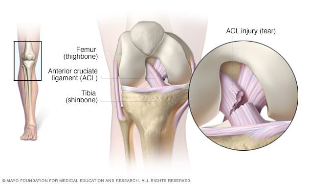
The anterior cruciate ligament is a ligament located at the level of the knee joint.
Anatomy of the anterior cruciate ligament
Position. The anterior cruciate ligament is located at the knee joint, which involves three bones: the femur which corresponds to the thigh bone; the tibia which designates the bone of the leg; and the kneecap, or patella, which makes up the knee bone. These three bones form three joints (1):
- The intermediate or patellofemoral joint, located between the kneecap and the lower part of the femur;
- The medial or femoro-tibial joint, located between the condyle of the femur, lower end of the femur, and the medial meniscus of the tibia, upper end of the tibia;
- The lateral or femoro-tibial joint, located between the condyle of the femur, the lower end of the femur, and the lateral meniscus of the tibia, the upper end of the tibia.
Two cruciate ligaments are located at the level of the double femoro-tibial joint and inside the joint capsule. The anterior and posterior cruciate ligaments form an X.
Structure. The anterior cruciate ligament is made up of fibrous connective tissue that has the particularity of being resistant (1).
Insertions. The anterior cruciate ligament is inserted at the level of the lateral condyle of the femur. Within the joint, it goes obliquely to attach itself to the anterior part of the tibia (1).
Knee joint
The anterior cruciate ligament plays an important role in the knee joint. It is particularly involved in the stability of the joint. During extension movements of the leg, the anterior cruciate ligament stretches and prevents the femur from sliding backwards in relation to the tibia (2). It thus makes it possible to oppose the hyperextension of the knee.
Pathologies of the anterior cruciate ligament
Some knee pain may be related to anterior cruciate ligament damage.
Sprain. This problem corresponds to an elongation or a tear of the ligaments. It can especially occur in the anterior cruciate ligament. A sprain is manifested by sharp pain and can prevent certain movements (1).
Cruciate ligament rupture. The rupture of the anterior cruciate ligament is a fairly common problem in athletes. The rupture of this ligament causes phases of dislocation of the knee as well as intense pain (3).
Knee dislocation. Usually linked to a violent shock, it corresponds to a loss of adhesion in a knee joint. Dislocations are often accompanied by inflammation, sprain or joint immobility (1).
Treatments
Medical treatment. Depending on the pathology diagnosed, certain drugs may be prescribed to reduce pain and inflammation.
Surgical treatment. Depending on the pathology diagnosed, a surgical intervention can be carried out with, for example, the reconstruction of the anterior cruciate ligament in the event of rupture.
- Arthroscopy. This surgical technique allows the joints to be observed and operated on. It is commonly used for the knee joints.
Physical treatment. Physical therapies, through specific exercise programs, are often prescribed such as physiotherapy or physiotherapy.
Examination of the anterior cruciate ligament
Physical examination. First, a clinical examination is performed to identify painful movements.
Medical imaging exam. Radiography, ultrasound or MRI can be used to assess pain in the knee.
History
In 2013, two surgeons from the University of Medicine of Louvain in Belgium published in the journal Journal of Anatomy, new results suggesting the presence of another ligament within the knee: the anterolateral ligament (4) (5 ). The presence of the latter had been suggested as early as 1879 by Dr Paul Segond, French surgeon. During his work on “knee blood effusions”, the researcher noted the existence of a “pearly band” (6).