Contents
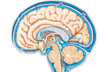
The term “hydrocephalus” consists of two words, literally translated from Latin as “water” and “brain”. With this disease, an excessive amount of cerebrospinal fluid – cerebrospinal fluid – is formed in the brain. This fluid serves as a shock absorber that protects the brain from physical influences, carries nutrients, and removes metabolic products. If the cerebrospinal fluid is formed in excess, the intracranial fluid pressure on the brain structures increases. Negative symptoms of the disease significantly worsen the quality of life of the patient. Moderate hydrocephalus is one of the varieties of this pathology.
Classification of hydrocephalus

The disease takes the following forms:
Moderate internal hydrocephalus – the accumulation of cerebrospinal fluid occurs in the ventricles of the brain;
Moderate external hydrocephalus – CSF accumulates in the subarachnoid space;
Moderate mixed hydrocephalus – combines the symptoms of both of the above varieties of the disease;
Moderate replacement hydrocephalus – cerebrospinal fluid replaces the atrophying substance of the brain, is more often diagnosed in old age, may accompany Alzheimer’s disease.
Any form of moderate hydrocephalus can be either congenital or acquired. Congenital moderate hydrocephalus is the result of birth trauma, infections suffered by the child during fetal development. The acquired form of the disease appears as a complication of somatic diseases, as well as as a result of a traumatic brain injury.
What leads to the development of the disease

Moderate hydrocephalus is based on impaired outflow of cerebrospinal fluid into the canal of the spinal cord, where it is normally absorbed into the venous network of the circulatory system.
Factors leading to the appearance of the disease:
Arterial hypertension;
Stroke;
Atherosclerosis;
A cyst or tumor that compresses the ventricle and stands in the way of CSF circulation;
Osteochondrosis;
Hernia of the cervical spine;
Neuroinfections – meningitis, encephalitis;
Alcoholism;
Craniocerebral injuries caused by bruises of the head during a fall from a height, impact, car accident.
Symptoms and manifestations of the disease

for a long time, the main and only symptom of the disease may be headaches, which appear most often in the morning. this symptom is due to a long stay in a horizontal position. headaches, as well as increased intracranial pressure, at the beginning of the development of the disease may not be. often hydrocephalus is detected only when diagnosing the state of the brain for a completely different reason.
sooner or later, an excessive amount of cerebrospinal fluid still manifests itself as signs of a violation of the functional capabilities of the brain, oxygen starvation.
untreated moderate hydrocephalus can lead to stroke, intellectual impairment.
symptoms of the disease in the development stage:
Visual and hearing impairments;
Loss of memory, inability to perform many intellectual operations;
Violation of fine motor skills, change in gait;
Violation of concentration;
Increased fatigue;
Insomnia, sleep disturbances;
Irritability;
Loss of the ability to navigate in space.
In the later stages of the development of the disease, vomiting, urinary incontinence, and cerebral edema develop. These symptoms are a consequence of a complete blockage of the outflow of cerebrospinal fluid, the so-called occlusive crisis.
Diagnosis of moderate hydrocephalus

The most reliable data on the state of the brain, on the anatomy of the internal cavity of the cranium, on the amount of cerebrospinal fluid and changes in brain structures due to increased intracranial pressure are obtained using MRI (magnetic resonance therapy). An alternative to this study is an x-ray of the skull in two projections.
Additional diagnostic methods:
Angiography – an x-ray with a contrast agent, which allows you to see stenosis of cerebral vessels, aortic aneurysm;
Ultrasound of the brain;
Examination of the fundus with the help of ophthalmoscopy, which allows to determine the presence or absence of edema of the optic nerve head;
Analysis of cerebrospinal fluid taken by lumbar puncture – allows you to detect pathogenic microorganisms in the infectious nature of the disease.
The tactics of the diagnostic examination is determined by the attending physician – a neurologist or neurosurgeon. You may need to consult an endocrinologist, psychotherapist, infectious disease specialist.
Medical treatment of moderate hydrocephalus

In the initial stage of the disease, it is possible to compensate for the symptoms of moderate hydrocephalus with the help of medications.
Most often assigned:
Diuretics to remove excess fluid from the body;
Preparations for the prevention of potassium and magnesium deficiency, excreted in significant quantities along with the liquid – Asparkam, Panangin;
Drugs to improve cerebral circulation;
Multivitamins and immunomodulators to strengthen the body’s defenses and accelerate recovery.
The appointment of a treatment regimen is carried out by the attending physician, self-administration of drugs can lead to serious complications.
Surgical treatment of moderate hydrocephalus

Although the prognosis of moderate hydrocephalus is quite favorable, with the development of symptoms of the disease, indications for surgical intervention may arise:
Severe headaches that are not relieved by taking analgesics;
Convulsions;
Intellectual impairment;
Movement coordination disorders;
Loss of control over bowel movements and urination.
The tactics of intervention depends on the cause of moderate hydrocephalus, the stage of the disease. Surgical Options:
Removal of a tumor or cyst that disrupts the circulation of cerebrospinal fluid;
Ventriculocisternoscopy endoscopy, which creates an artificial pathway for the removal of cerebrospinal fluid;
Shunting, which allows you to dump excess amounts of CSF.
Most often, shunting is performed – an operation worked out to the smallest detail several decades ago. A catheter is inserted into the cavity of the ventricle, the valve of which opens after reaching an increased level of intracranial pressure. After restoration of normal values, the valve closes.
The output of cerebrospinal fluid occurs in the body cavity of the patient, capable of transforming it and removing it from the body:
Abdominal cavity (preferred);
Atria;
gallbladder;
Large vessels;
Ureter.
If hydrocephalus is caused by infectious factors, shunting is not performed so as not to provoke inflammatory processes in the tissues of the patient’s body. Endoscopic intervention requires qualified specialists and modern equipment. In the bottom of the ventricle, overflowing with CSF, artificial openings are created for the withdrawal of cerebrospinal fluid into the basal cisterns in the occipital part of the brain.
In some cases, the removal of brain tumors of any etiology, cysts with helminth cysts, scars after a stroke can give a significant relief to the patient’s condition. If the formations do not affect large vessels, and have not grown through the tissues of the brain, the relief of the condition occurs very quickly.
In order not to aggravate the first negative symptoms of the disease, if you suspect moderate hydrocephalus, you should undergo a full examination.


