Contents
Hydrocephalus is a neurological disorder characterized by the production of excessive amounts of cerebrospinal fluid. This circumstance leads to an increase in craniocerebral pressure and other negative symptoms. Hydrocephalus can be congenital or acquired, it is classified on various grounds. Mixed hydrocephalus is one of the rare, but very dangerous types of dropsy of the brain.
Clinical picture of mixed hydrocephalus

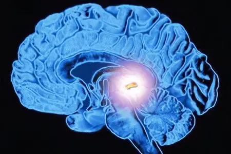
The brain is protected from external influences by several membranes. The space between them is called the subarachnoid space. Inside the brain there are peculiar cavities – the ventricles, in which the cerebrospinal fluid circulates. With a concentration of cerebrospinal fluid in the ventricles, the disease is referred to as an internal form of hydrocephalus, with the accumulation of cerebrospinal fluid in the subarachnoid cavity, external hydrocephalus is diagnosed.
If the cerebrospinal fluid overflows both cavities, the diagnosis is mixed hydrocephalus. At the same time, destruction of brain tissues is observed, the place of which is occupied by cerebrospinal fluid. Its excess compresses the structures of the brain, which is manifested by intellectual impairment, memory loss, pain syndrome. Most often, such complications appear in old age due to brain atrophy.
Mixed atrophy can appear against the background of alcoholism, the consequences of a traumatic brain injury, and severe atherosclerosis. In severe cases, during the development of the disease, the patient falls into a coma, he may have a fatal outcome, or there will be prerequisites for disability. Lack of muscle control leads to involuntary urination and defecation. The patient loses the ability to self-service, cannot do without outside help.
Causes of mixed hydrocephalus

Hydrocephalus a few years ago was considered a pathology inherent in childhood. Until now, the lion’s share of diagnosed cases of mixed hydrocephalus occur in the first six months of a child’s life, or in the period of intrauterine development.
Causes of the mixed form of hydrocephalus in children:
Birth injury;
Pathologies of intrauterine development;
Infection of the fetus, diseases transferred by a pregnant woman;
Fetal intoxication during pregnancy;
Genetically determined fetal pathologies.
Causes of mixed hydrocephalus in adults:
Spinal injuries and craniocerebral injuries;
Consequences of meningitis and neuroinfections;
Intoxication with alcohol, narcotic drugs;
Fragility of the cervical spine in old age;
Arterial hypertension;
Atherosclerosis.
Mixed hydrocephalus in childhood significantly inhibits the intellectual and mental development of the child. In adults, this disease may not manifest itself in the early stages, but over several years or months it significantly impairs the quality of life.
Symptoms of mixed hydrocephalus

The main symptoms are increased intracranial pressure and such neurological symptoms as impaired memory, speech, emotional reactions to the surrounding reality.
Symptoms in adults:
Nausea and vomiting in the morning;
Passivity, loss of interest in the environment;
Intense headaches;
Dislocation of the brain, or its displacement along the main axis;
Drowsiness, as a harbinger of deterioration;
Vision disorders.
With the development of mixed hydrocephalus, seizures of the epileptic type may begin, the ability to remember and manipulate numbers is lost. Answers to the simplest questions are difficult for the patient, he inadequately responds to the appeal to him.
Symptoms in children:
Bulging of the “font”;
Astigmatism;
Fits of crying with head thrown back;
Convulsions;
Omission of the eyeball (symptom of grefe);
Frequent restless crying;
Refusal to eat;
Decreased hearing and visual acuity;
Violation of coordination of movements.
The most noticeable visual symptom is an increase in the skull, especially noticeable in the frontal part. A slight increase in the frontal lobes can be recorded even in an adult. Mixed hydrocephalus, even in the most moderate form, requires immediate medical attention. If the necessary assistance is not provided in a timely manner, problems with reduced intelligence and the inability of the child to maintain independent life support are inevitable.
How is mixed hydrocephalus diagnosed?

The clinical picture of hydrocephalus usually indicates its presence so clearly that in most cases the doctor has no doubts about the diagnosis already during the initial examination. Additional studies are being conducted to clarify the type of hydrocephalus and the stage of its development.
There are “three whales”, or three main signs, on the basis of which the diagnosis of “mixed hydrocephalus” is made:
Symptoms;
MRI data;
Analysis of the condition of the fundus.
If all of them confirm the alleged diagnosis, it is put without any doubt. If there are doubts on at least one basis, or a firm answer is “no”, most likely, instead of hydrocephalus, the patient suffers from a pathology with similar symptoms.
Diagnosis of hydrocephalus:
X-ray of the skull – a discrepancy between the seams of the skull, thinning of its bones, on the inner surface of the cranial vault, a symptom of “finger impressions”, asymmetry of the skull are revealed;
Echoencelography, or ultrasound scan of the brain, determines the degree of intracranial pressure;
Ultrasonography – scanning the brain in infants through an open fontanel;
Ophthalmoscopy, perimetry, determination of visual acuity – an ophthalmologist determines the degree of visual impairment, the condition of the optic discs;
MRI of the brain – determines the type of hydrocephalus, the cause of the development of pathology (cyst, tumor), congenital anomaly;
Lumbar puncture with the study of cerebrospinal fluid – helps to identify the causes of the disease.
If mixed hydrocephalus is caused by an infection, PCR diagnostics is recommended to clarify the type of infectious factor.
Treatment of mixed hydrocephalus

The main treatment for this complex disease is surgery.
Conservative therapy, although used to reduce the volume of CSF production, is used only in special cases:
Mitigation of neurological symptoms in preparation for surgery;
Treatment of mild acquired mixed hydrocephalus resulting from ventricular hemorrhage, traumatic brain injury or alcoholism of the patient.
Most often, diuretics are used, which reduce the amount of fluid in the body and the volume of cerebrospinal fluid in the ventricles and subarachnoid space.
Groups of drugs for the treatment of mixed hydrocephalus:
Diuretics;
Nootropics and means to improve cerebral circulation;
Drugs that replenish potassium and magnesium, actively excreted along with diuretics – Panangin, Asparkam;
sedatives;
Venotonics;
Fortifying drugs.
In difficult cases, an operation is performed to install a shunt between the subarachnoid cavity, the ventricles of the brain and other cavities of the patient’s body to reduce intracranial pressure.
External shunting routes – where the shunt is taken from the brain:
into the peritoneum;
In the heart;
In the lungs;
In a vein;
In the urethra.
Methods of internal shunting:
Creating a message between the ventricle of the brain and the occipital cistern (Thorkildsen’s operation);
Creating a message between the ventricle of the brain and the interpeduncular cistern in the region of the gray tubercle;
Implantation of stents that expand the circulation of CSF;
Expansion of the lumen of the aqueduct of the brain;
Creating an opening between the ventricles for CSF circulation.
The introduction of a shunt into the brain requires regular monitoring of the patient by a neurosurgeon, a neurologist. If a shunt is placed in a child, as the head grows, a second operation is required to replace it with a larger shunt. In case of infection of an artificially created canal, courses of antibiotic therapy are prescribed.


