


Contents
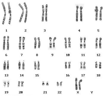
The karyotype is a set of chromosomes that is present in all somatic cells (that is, those that are not sex cells) in the human body. The structure of the chromosome depends on both species and sex. In humans, the karyotype is made up of the sex-determining autosomes and chromosomes. Autosomes are found in both female and male patients. Every healthy person has 22 pairs of autosomes. The X and Y chromosomes determine the type of sex. The female has two X chromosomes, while the male has one of both X and Y chromosomes. Changes or abnormalities in this structure are called chromosomal abnormalities. Depending on the type of mutation, there are: deletions, inversions and translocations. In cases where the number of chromosomes has changed, it is called trisomy.
Karyotype examination – indications
Disturbances in the structure chromosomes can contribute to the development of genetic defects, genetic diseasesand also be the reason miscarriages. It also happens that they prevent a woman from getting pregnant or are a source of great difficulty in maintaining it. Karyotype examinationthat allows you to analyze chromosomes inside the cells and thus to detect possible abnormalities in their number or shape, it should be performed in both a woman and a man. It is recommended to conduct it, first of all, in the situation in which it occurred spontaneous miscarriageespecially if it was the first time. It should also be done in the case of a woman she had a miscarriage more than once or has given birth to a stillborn child.
Research karyotype should also be considered by those who are planning to start a family and have had family history of the disease genetic diseases. Research allows you to determine whether the child will be a carrier of the mutation responsible for a given disease.
Karyotype disorders and miscarriage
For women who have difficulty carrying a pregnancy or had a stillborn baby, the likelihood of abnormal build or mutation is likely chromosomes is about 16 percent. Karyotype examination is a cytogenetic test. Miscarriage in the first trimester of pregnancy may most likely mean that it is caused by a mutation within chromosomes. It is estimated that such defects appear in approximately 2–4 percent of cases.
Za miscarriages most often correspond to the so-called balanced translocation or Robertson translocation that is based on fragment displacement chromosome to another place to the same or another chromosome.
A balanced translocation occurs when the amount of genetic material has not changed, but only the location in the genome. If such a mutation occurs in one of the parents, the child may develop an unbalanced translocation, i.e. the amount of genetic material changes. Consequently, fetal death very often occurs and miscarriage. A Robertson translocation is a combination of the long arms chromosomes, Co examination of the karyotype shows as missing one chromosome.
He is very often responsible for the problem with carrying and maintaining a pregnancy mosaicism (especially when it occurs in women). Mosaicism is the presence of more than one lineage in a human (for example, instead of 46XX chromosomes all somatic cells show a combination of 46XX and 45X cells). If one is missing the X chromosome, there is talk of Turner syndrome. Research shows that 50 percent of women struggle with it genetic disease it comes to miscarriages.
Karyotype – the course of the study
To complete karyotype testing it is enough to take a small amount of blood from the patient. There is no need to prepare for it and you can take a sample at any time. Typically, genetic material from leukocytes, i.e. white blood cells, is analyzed. In situations where, for example, during an ultrasound examination of a pregnant woman, the doctor noticed something disturbing or the woman lost a previous pregnancy or gave birth to a stillborn child, the material for the examination is the amniotic fluid or the chorionic fragment.
Carrying out a cytogenetic test requires culturing (using an in vitro procedure) a certain number of cells, as a result of which it takes about three weeks for the result to be obtained. After the cells are grown, substances are added to them that stop cell division at a certain stage (usually a metaphase in which chromosomes are best visible). Later, preparations are made chromosomowe being the basis for the assessment of the number and morphology of individual chromosomes. The test description provides a graphical listing of all chromosomes the patient, as well as a detailed description containing information on whether they are corrector not.