Contents
Fertilization: how do you get pregnant?
Without fertilization, no pregnancy! But when is the fertile period really? How does fertilization take place and how does the embryo evolve in the hours that follow it? Decryption of this key moment that is the meeting between the sperm and the egg.
The fertility period
Fertilization describes the meeting and fusion between the male gamete, the sperm, and the female gamete, the oocyte. More precisely, it is considered to be completely finished when the genetic information of the two gametes unites to form a new cell, the genesis of the embryo.
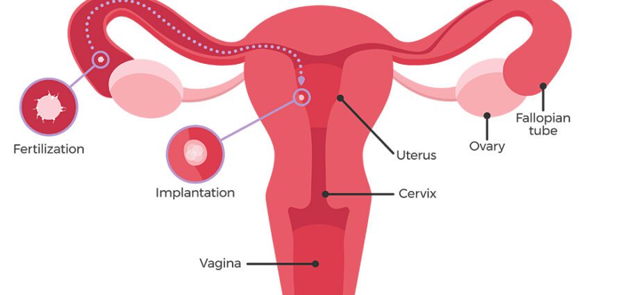
So that the woman can become pregnant (in the case of a spontaneous pregnancy), this meeting is only possible in period of fertility, that is to say during ovulation. Indeed, under the effect of the physiological hormonal impregnation of the woman and more particularly estrogen, only one (more rarely two) oocytes arrive (s) each month at maturity: the follicle of De Graaf. Under the influence of the hormone LH (luteinizing hormone), it is expelled from the ovary around the 14th day of the cycle. Between 12 and 24 hours later, the oocyte is intercepted by the fallopian tube. He then “waits” for a few hours in the tubal bulb (the part of the tube furthest from the uterus) with a view to possible fertilization. Moreover, if this takes place, the first cells of the embryo begin their development in the said bulb, before migrating to the endometrium, 2 to 3 days later.
Man, for his part, does not know this calendar imperative for fertilization. Unlike the woman whose oocyte production is in a way predefined by the ovarian reserve and then her menstrual rhythm, the man continually renews his “stock” of sperm from puberty, during a cycle of 64 days. It is spermatogenesis. To promote fertilization, however, the spermatozoon must be mature, mobile and of a typical shape, ie composed of:
- of a head, itself subdivided into a nucleus, carrying the 23 paternal chromosomes and an acrosome, anterior part of the gamete’s head, a kind of small bag which will release the enzymes necessary for the acrosomal reaction and therefore for the penetration of the spermatozoon into the ‘ovum,
- an intermediate piece,
- of a scourge, guaranteeing the oscillations necessary for the movement of the spermatozoon in the female tract.
The path of the sperm to the egg
At the time of ejaculation, between 2 and 5 cm3 of semen reaches the vagina, or between 60 and 500 million sperm. Among them, only 100 to 200 gametes manage to travel the 13 to 15 cm which separate the vagina from the tubal bulb where the ovum is housed. They then go through different stages:
Passage of the cervix
Surrounded by seminal fluid which constitutes a kind of alkaline buffer, the sperm are protected from the acidity of the vagina. However, this protection is insufficient in the majority of cases and a large part of the sperm die before they have even crossed the cervix.
Once the cervix is reached, the “surviving” spermatozoa evolve in an environment more conducive to their survival. Indeed, during ovulation, the properties of the cervix and the mucus that lines it change. The cervix relaxes, the canal between the vagina and the uterus widens, and the mucus, accumulated in a plug to prevent the passage of sperm outside the period of ovulation, becomes permeable, more liquid. The environment hitherto unfriendly to spermatozoa thus allows their passage. Moreover, some spermatozoa can survive at this stage for up to 3 or 4 days after intercourse. This explains why sometimes fertilization is not immediate!
During this first stage and throughout its few (potential) days of life in the female tract, the sperm undergoes physiological changes. We then speak of capacitation. During capacitation, the cell membrane of male gametes evolves. They then acquire a new motility, called hyperactivity. The objective: to allow them, at the time of fertilization, to carry out the acrosomal reaction, that is to say the release of enzymes necessary for the connection between the spermatozoon and the zona pellucida (external) of the oocyte.
The penetration of cumulus oophorus cells
Housed in the tubal bulb, the oocyte is surrounded by cells of the cumulus oophorus, which before ovulation, attached the heart of the ovum (the granulosa) to the antrum, a cavity of follicular fluid necessary for its expulsion from the ovary. During fertilization, the acrosomal reaction allows the passage of the sperm through these cumulus cells. Thanks to its new hypermobility, the spermatozoon then makes its way to the oocyte.
The meeting between the egg and the sperm
It is also thanks to the acrosomal reaction that the meeting and the fusion between the oocyte and the spermatozoon is possible. Indeed, thanks to the release of certain enzymes (hyaluronidase, acrosine), the spermatozoon can penetrate the zona pellucida before attaching itself to the membrane of the oocyte, its inner layer. In parallel, still under the effect of this reaction, the envelope of the oocyte relaxes to facilitate the passage of said sperm.
Once the sperm is secured, several physiological processes take place to increase the chances of getting pregnant:
- The pellucid area hardens again to prevent the passage of new sperm and provide protection for the future embryo,
- the membranes of the oocyte and sperm merge.
After this fusion, the nucleus of the sperm begins to grow. It contains the genetic heritage that the father transmits to his child, namely 22 chromosomes and an X or Y chromosome which will define the sex of the future baby. At the same time, the nucleus of the egg, which also contains 22 maternal chromosomes and one X chromosome, increases in size. The two (pro) nuclei are then found in the center of the oocyte where they merge, associating the 46 chromosomes of the unborn child. It is the zygote, the very first cell in the embryo, which already has its own genome. The fertilized oocyte has only a few hours and yet all the physical characteristics, the fundamental traits of the child are already programmed.
After fertilization: segmentation …
Between 22 and 26 hours after fertilization, the chromosomes are distributed in the first two constituent cells of the embryo: blastomeres. Cell division, or segmentation, begins, to continue, in these first days of the embryo’s life, at very high speed:
- Around the 50th hour, the blastomeres divide into 4,
- Around the 60th hour, these same cells divide into 8,
- When the embryo reaches the 16-cell stage (and up to the sixth cell division), it is described by the term morula, in reference to its blackberry-like appearance. 72 hours after fertilization, the morula begins its journey from the tube to the uterine cavity. For the next 3 days, cell division continues until the embryo has 64 cells. The morula, which until then was the original size of the oocyte, begins to grow.
… and the formation of the embryo
Around the 4th day, the morula becomes blastocyste. At this stage of its development, it consists of two types of cells. In its center, larger cells that will form the embryonic button. At the periphery, the cells correspond to the trophoblast, the future placenta. Once the migration to the body from the uterus is complete, the zona pellucida from the old oocyte disappears, allowing the embryonic pole to attach to the endometrium. Between the 7 and 10 th days after fertilization, the implantation takes place: well sheltered in the uterine lining, the blastocyst can continue to develop.
During the 2 nd and 3 rd weeks of pregnancy, the embryonic button evolves and takes the form of a disc first formed of two (the inner and outer layers), then 3 layers of cells. Between the 15th and 17th day after fertilization, the outer layer thickens to reveal the outline of the head and tail of the embryo. Then gradually, from the three layers of the embryonic disc will flow all the other cells of the embryo, to give birth, a few months later, to the baby.


