


Contents
Bladder scanner: what is it for?
The bladder scanner, or bladder ultrasound machine, is a portable, battery-operated device intended to measure the volume of the bladder with precision thanks to its ultrasound technology. This is because it uses high-frequency, low-energy sound waves which are reflected by the walls of the bladder. No probe or other medical instrument needs to be introduced inside the body. The measurement of the bladder volume by this method makes it possible to evaluate the urinary retention, to find a possible post-voiding residue and to avoid the catheters which frequently lead to urinary infections in the patients. It does not cause trauma or pain.
What is a bladder scanner?
The bladder scanner, or bladder ultrasound, is a non-invasive ultrasound device that assesses the bladder filling rate, for patients who are unable to feel or express their own needs, using advanced scanning technology. Battery powered, it is easy to use by trained caregivers and quickly provides the information they need.
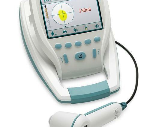
The device is made up of two interconnected parts:
- a portable probe, placed on the abdomen towards the bladder after application of an ultrasound conducting gel to the patient’s skin in the suprapubic area;
- a base unit incorporating a display screen, having a sighting circle appearing on the image of the scanned bladder in order to assist the operator.
The bladder scanner:
- measures the reflection of ultrasound in several planes inside the abdomen differentiating the bladder from surrounding tissues;
- provides cross-sectional images of the bladder;
- and using a microprocessor, calculates and displays, on a liquid crystal display, the patient’s bladder volume, in ml.
The information measured by the bladder scanner can be printed using an integrated printer or transmitted to a computer for review, printing or archiving.
What is a bladder scanner used for?
Bladder scan measurement can:
- substitute for diagnostic probing to accurately assess bladder volume, bladder emptying time and measure post-void residual volume;
- help to avoid a number of unnecessary catheters which can be painful and can lead to urethral trauma and urinary tract infections.
The use of the bladder scanner is recommended:
- postoperatively: after general anesthesia, urinary retention is a complication associated with over-distension of the bladder that can lead to the risk of permanent damage to the detrusor muscle of the bladder;
- in urology: the measurement of the bladder volume is a necessity for exploring voiding disorders, evaluating the post-voiding residue, particularly in surgery for functional disorders of the lower apparatus, in the presence of benign prostatic hypertrophy and in cases of incontinence or ‘repeated urinary tract infections, or to confirm the existence of a bladder in patients who are sometimes difficult to examine;
- in neurology: central and peripheral neurological damage preventing normal emptying of the bladder results in urinary retention or incontinence, partial emptying of the bladder, over-distension of the bladder, kidney stones, increased spasticity and autonomic reflex disorders;
- in geriatrics: the elderly are among the people most exposed to the risk of urinary retention, in particular due to physiological changes associated with aging, the use of anticholinergic drugs, cognitive disorders or even diabetes. They also usually suffer from infections due to their advanced age if conventional probing techniques are used.
The use of the bladder scanner is also possible to supplement other analyzes and auscultations within the framework of the study of the size of the bladder. These examinations make it possible to know the volume of the organ and to understand its functioning or on the contrary the dysfunctions.
Finally, use you browse the scanner can be extended to biofeedback programs, control training and bladder continence.
How is a bladder scanner used?
The bladder scanner is easy to use, fast, non-invasive and comfortable.
HOW TO USE
- comfortably place the patient flat on their back;
- partially uncover the patient, revealing only the lower abdomen and covering the rest of the body;
- make sure that the device is correctly configured (date and time), plug it in, set it to “on”, to check that it works;
- turn on the screen;
- press the “scan” button;
- select the patient’s sex;
- use the “male” selection if the patient is a female who has undergone a hysterectomy;
- clean the rounded end of the probe head by wiping it carefully with a swab soaked in aqueous chlorhexidine;
- apply an appropriate amount of ultrasound gel to the patient’s abdomen;
- smooth the gel to remove any air bubbles that may block the transmission of ultrasound;
- place the probe head approximately 3 cm from the pubic symphysis (i.e. the articulation that unites the two pubic bones in the middle position) pointing to the foreseeable location of the bladder;
- orient the probe slightly downwards towards the tailbone;
- press the scan button on the probe to start the scan or press the scan button on the software;
- the screen displays the largest volume measured, the status of the instrument, the measurement and the aiming target;
- if the reticle is not centered on the bladder, adjust the probe and repeat the gesture until centering is obtained;
- when the aim is correct, press the “done” button. The results screen is then displayed;
- print the results.
Precautions for use
- clean the screen;
- disinfect the probe head before and after use to avoid infectious and bacterial contaminations;
- check that there is sufficient paper in the machine;
- also check that the bladder scanner battery is fully charged before the examination;
- store the device in a dry place.
Potential adverse effects and contraindications
A patient with urinary incontinence may be inconvenienced by the pressure exerted by the catheter. Some potential unwanted effects may also occur with the use of the bladder scanner, such as skin irritation or an allergic reaction to the ultrasound conductive gel. Finally, certain clinical conditions are likely to interfere with the examination and cause measurement errors:
- obesity;
- abdominal scars in the bladder area;
- ovarian cysts;
- changes in the shape or position of the bladder after surgery or linked to the presence of stones;
- clots in the bladder;
- fluids contained in the pelvic cavities;
- pregnancy ;
- muscle spasms ;
- abdominal hernias.
How to choose a bladder scanner?
For several years, portable bladder ultrasound systems have been the subject of significant technological developments.
For users wishing to have all-in-1 stand-alone equipment, it is advisable to opt for a complete version with integrated monitor and printer, particularly suitable for establishments such as nursing homes or retirement homes. The image captured by the probe can be viewed in real time on the screen of the chosen digital medium (tablet, PC or monitor), which allows medical workers to be able to reposition the probe instantly in the most suitable angle for the measurement in order to obtain the most reliable data possible.
Finally, the bladder scanner can be supplied in a bag, in order to transport it in complete safety.