


Contents
Intestinal dysfunction is a common symptom of many diseases. Many of these ailments are not even contagious. Since diarrhea accompanies most infectious diseases, it may seem strange that bovine viral diarrhea is not a symptom, but a separate disease. Moreover, with this disease, bowel dysfunction is not the main symptom.
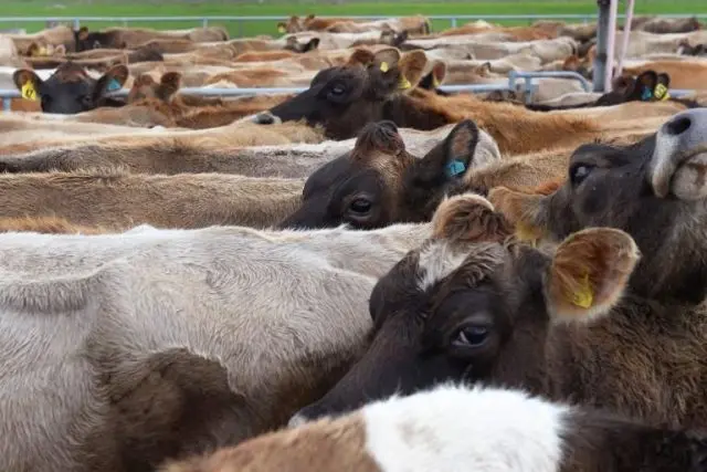
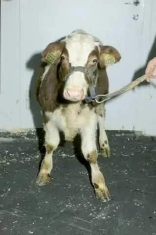
Highly contagious viral disease. Diarrhea is the lesser of the evils that characterize this disease. With viral diarrhea, the mucous surfaces of the intestines, mouth, tongue, and even the nasolabial mirror become inflamed and ulcerated. Conjunctivitis, rhinitis and lameness develop. Fever appears.
The disease causes great economic damage on farms, as sick pregnant cows are aborted, and lactating cows reduce milk yield. Viral diarrhea is widespread throughout the world. Only strains of the virus may differ.

Causative agent
The causative agent of this viral disease in cows belongs to the genus pestiviruses. At one time, it was believed that this type of virus could be carried by blood-sucking insects and ticks, but later it was found that viral diarrhea of cows was not transmitted in this way.
There are 2 genotypes of viruses that cause infectious diarrhea in cows, but they do not differ in virulence. Viruses with the BVDV-1 genotype were previously thought to cause milder disease than BVDV-2. More recent studies have not confirmed this. The only difference is that type XNUMX viruses are less common in the world.
The diarrhea virus is very resistant to low temperatures in the external environment. At -20 °C and below, it can persist for years. In the pathological material at -15 ° C, it persists for up to 6 months.
The virus is not easy to “finish off” even at positive temperatures. It withstands + 25 ° C during the day without reducing activity. At + 35 °C it remains active for 3 days. Bovine diarrhea virus is inactivated only at +56°C and after 35 minutes at this temperature. At the same time, there is an assumption about the presence of heat-resistant strains of viral diarrhea.
The virus is sensitive to disinfectants:
- trypsin;
- ether;
- chloroform;
- deoxycholate.
But not all is well here either. Ether-resistant strains of viral diarrhea also exist, according to Hack and Taylor’s research.
An acidic environment is capable of “finishing off” the virus. At pH 3,0, the pathogen dies within 4 hours. But in excrement can be stored up to 5 months.
Due to such “quirkiness” of the causative agent of viral diarrhea, today, according to various sources, from 70 to 100% of the total number of cows in the world is infected with this disease or was ill earlier.
Sources and routes of infection
Viral diarrhea is transmitted in several ways:
- direct contact of a sick cow with a healthy animal;
- intrauterine infection;
- sexual transmission even with artificial insemination;
- blood-sucking insects;
- when reusing nasal forceps, needles or rectal gloves.
It is almost impossible to avoid contact of sick cows with a healthy herd. There are always up to 2% of infected animals in the herd. The reason for this is another way of spreading the infection: intrauterine.
Due to the latent course of the disease, many cows are able to calve with an already infected calf. A similar situation occurs if there was an outbreak of an acute form of the disease in the early stages of pregnancy. The body of a calf, infected in the womb, recognizes the virus as “its own” and does not fight it. Such an animal sheds the virus in large quantities throughout its life, but does not show signs of illness. This feature contributes to the “success” of cow viral diarrhea among other diseases.
Since latently ill bulls and producers with an acute form of the disease shed the virus along with sperm, cows can become infected during artificial insemination. Freezing semen in liquid nitrogen only contributes to the persistence of the virus in the seed. In the body of cattle producers, the virus persists in the testes even after treatment. This means that a bull that has been ill and treated still remains a carrier of the cow diarrhea virus.
The virus is also transmitted through the blood. These are the unsterilized instruments that are already familiar to everyone, reusable needles of syringes or the reuse of reusable ones and the transmission of the virus by blood-sucking insects and mites.
The usual duration of the incubation period is 6-9 days. There may be cases when the incubation period lasts only 2 days, and sometimes stretches up to 2 weeks. The most common clinical signs of viral diarrhea include:
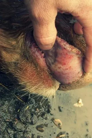
- ulceration of the mouth and nose;
- diarrhea;
- high fever;
- lethargy;
- loss of appetite;
- reduction in milk yield.
But the symptoms are often blurred or poorly expressed. With insufficient attention, the disease is easy to miss.
The general set of symptoms that can be with viral diarrhea:
- heat;
- tachycardia;
- leukopenia;
- depression;
- serous discharge from the nose;
- mucopurulent discharge from the nasal cavity;
- cough;
- salivation;
- lacrimation;
- catarrhal conjunctivitis;
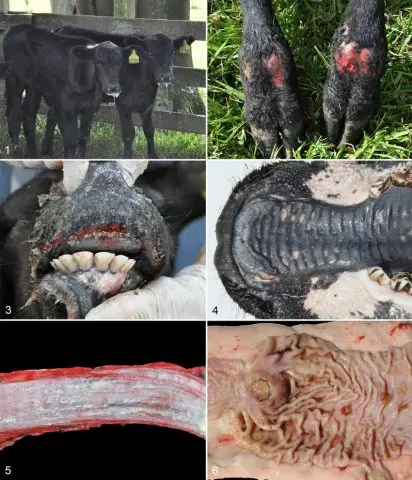
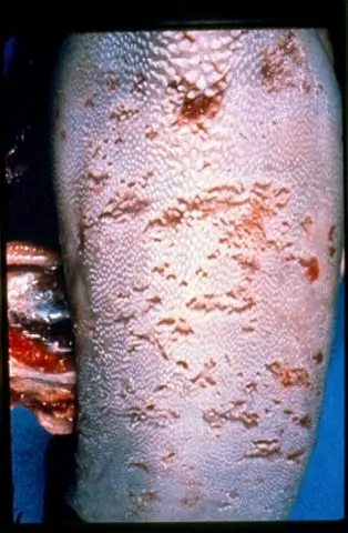
- erosion and ulcers on any mucous membranes and in the interhoof gap;
- diarrhea;
- anorexia;
- abortions in pregnant cows.
The specific set of symptoms depends on the type of course of the disease. Not all of these signs of viral diarrhea are present at the same time.
Course of the disease
The clinical picture is diverse and largely depends on the nature of the course of viral diarrhea:
- acute;
- subacute;
- chronic;
- latent.
The course of the acute form of the disease varies depending on the condition of the cow: pregnant or not.
Acute current
In an acute course, symptoms appear suddenly:
- temperature 39,5-42,4 ° C;
- depression;
- refusal to feed;
- tachycardia;
- frequent pulse.
After 12-48 hours, the temperature drops to normal. Serous discharge from the nose appears, later becoming mucous or purulent-mucous. Some cows have a dry, hard cough.
In a severe form of an acute course, the muzzle of a cow may become covered with dried secretions. Further, foci of erosion can form under dry crusts.
In addition, viscous saliva hanging from the mouth is observed in cows. Catarrhal conjunctivitis develops with severe lacrimation, which may be accompanied by clouding of the cornea.
Round or oval foci of erosion with sharply defined edges appear on the mucous membranes of the oral cavity and the nasolabial mirror.
Sometimes the main symptom of viral diarrhea is lameness of cows, resulting from inflammation of the cartilage of the limb. It is not uncommon for cows to lame throughout the illness and after recovery. In isolated cases, lesions appear in the interhoof gap, which is why viral diarrhea can be confused with foot and mouth disease.
During a fever, the manure has a normal shape, but it contains mucous and blood clots. Diarrhea occurs only after a few days, but no longer stops until recovery. Manure is fetid, liquid, bubbling.
Diarrhea causes the body to become dehydrated. With a long course, the cow’s skin becomes hard, wrinkled and covered with dandruff. Foci of erosion and crusts of dried exudate appear in the inguinal region.
Affected cows can lose up to 25% of body weight within a month. Milk yields of cows are reduced, abortions are possible.
Acute course: non-pregnant cattle
In young cows with strong immunity, viral diarrhea in 70-90% of cases is almost asymptomatic. With careful observation, you can notice a slight increase in temperature, mild agalactia and leukopenia.
Young calves aged 6-12 months are very susceptible to the disease. In this category of young animals, the circulation of the virus in the blood begins from the 5th day after infection and lasts up to 15 days.
Diarrhea in this case is not the main symptom of the disease. More common clinical signs include:
- anorexia;
- depression;
- reduction in milk yield;
- nasal discharge;
- rapid breathing;
- oral injury.
Non-pregnant cows with an acute course of the disease shed less virus than those infected in utero. Antibodies begin to be produced 2-4 weeks after infection and persist for many years after the disappearance of clinical signs.
Previously, viral diarrhea in non-pregnant cows proceeded in mild forms, but since the late 1980s, strains have appeared on the North American continent that cause severe diarrhea.
Severe forms were characterized by acute onset of diarrhea and hyperthermia, sometimes leading to death. A severe form of the disease is caused by genotype 2 viruses. Initially, severe forms were found only on the American continent, but were later described in Europe. Viral diarrhea of the second type is characterized by a hemorrhagic syndrome, which leads to internal and external hemorrhages, as well as nosebleeds.
A severe form of the disease is also possible with a type 1 infection mutation. In this case, the symptoms are:
- heat;
- mouth ulcers;
- eruptive lesions of interdigital clefts and coronary region;
- diarrhea;
- dehydration;
- leukopenia;
- thrombocytopenia.
The latter can lead to petechial hemorrhages in the region of the conjunctiva, sclera, oral mucosa and vulva. In addition, after injections, prolonged bleeding from the puncture site is observed.
Acute course: pregnant cows
During pregnancy, a cow shows the same signs as a single animal. The main problem of the disease during pregnancy is infection of the fetus. The causative agent of viral diarrhea is able to cross the placenta.
When infected during insemination, fertility decreases and the percentage of early death of embryos increases.
Infection in the first 50-100 days can lead to the death of the embryo, while the expulsion of the fetus will occur only after a few months. If the infected embryo does not die within the first 120 days, then a calf is born with congenital viral diarrhea.
Infection between 100 and 150 days leads to birth defects in calves:
- the thymus;
- eye;
- cerebellum.
Tremors are observed in calves with cerebellar hypoplasia. They cannot stand. With eye defects, blindness and cataracts are possible. With the localization of the virus in the vascular endothelium, edema, hypoxia and cellular degeneration are possible. The birth of weak and short calves can also be caused by infection with viral diarrhea in the second trimester of pregnancy.
Infection in the period of 180-200 days causes a reaction of an already fully developed immune system. In this case, calves are born outwardly completely healthy, but with a seropositive reaction.

Subacute course
A subacute course with inattention or a very large herd can even be missed, since the clinical signs appear rather weakly, only at the onset of the disease and for a short time:
- temperature increase by 1-2 °C;
- rapid pulse;
- frequent shallow breathing;
- reluctant eating of food or complete refusal of food;
- short-term diarrhea within 12-24 hours;
- slight damage to the oral mucosa;
- cough;
- nasal discharge.
Some of these signs can be mistaken for mild poisoning or stomatitis.
In the subacute course, there were cases when viral diarrhea proceeded with fever and leukopenia, but without diarrhea and ulcers on the mucous membranes of the oral cavity. Also, the disease can occur with other symptoms:
- cyanosis of the mucous membranes of the mouth and nose;
- pinpoint hemorrhages on the mucous membranes;
- diarrhea;
- fever;
- atony.
Viral diarrhea was also described, which lasted only 2-4 days and was expressed in diarrhea and a decrease in milk yield.
chronic course
In the chronic form, the symptoms of the disease develop slowly. Cows are gradually losing weight. There is intermittent or persistent diarrhea. Sometimes even diarrhea may be absent. The rest of the signs do not appear at all. The disease can last up to 6 months and usually ends in the death of the animal.
Chronic diarrhea occurs in cows that are kept in improper conditions:
- poor feeding;
- unsatisfactory conditions of detention;
- helminthiasis.
Also, outbreaks of the chronic form of the disease are present in farms where an acute form of diarrhea was previously recorded.
latent flow
There are no clinical signs. The fact of the disease is established by a blood test for antibodies. Often, antibodies to this viral disease are found even in clinically healthy cows from farms where diarrhea has never been recorded.
Mucosal disease
It can be taken out as a separate form of the disease, which affects young animals aged 6 to 18 months. Inevitably leads to death.
The duration of this type of diarrhea is from several days to several weeks. It begins with depression, fever and weakness. The calf loses its appetite. Gradually, emaciation sets in, accompanied by foul-smelling, watery, and sometimes bloody diarrhea. Due to severe diarrhea, the body of the calf is dehydrated.
The name of this form comes from ulcers localized on the mucous membranes of the mouth, nose and eyes. With a strong lesion of the mucous membranes in young cows, strong lacrimation, salivation and nasal discharge are observed. Also, lesions can be in the interdigital gap and on the corolla. Because of them, the cow stops walking and dies.
This form of the disease occurs in prenatally infected young animals as a result of the “imposition” of its own virus on an antigenically similar strain of the pathogen from another sick individual.
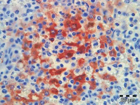
Diagnostics
The diagnosis is made on the basis of clinical data and the epizootic situation in the area. The final and accurate diagnosis is made after the study of pathological material. The virus isolated from the mucous membranes is differentiated from pathogens of other diseases that have similar symptoms:
- fungal stomatitis;
- foot and mouth disease;
- infectious ulcerative stomatitis;
- rinderpest;
- parainfluenza-3;
- poisoning;
- malignant catarrhal fever;
- paratuberculosis;
- eimeriosis;
- necrobacteriosis;
- infectious rhinotracheitis;
- mixed alimentary and respiratory infections.
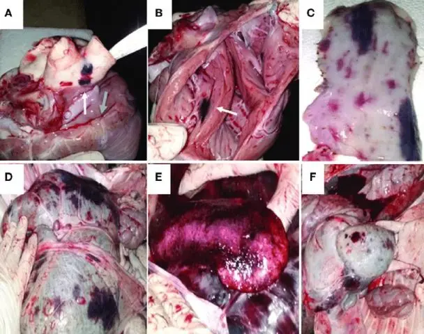
For pathological studies, parts are selected where the erosion of the mucous membranes is most pronounced. Such changes can be found on the gastrointestinal tract, lips, tongue, nasal mirror. In the intestine, sometimes there are extensive foci of necrosis.
The respiratory system is less affected by viral diarrhea. Erosion is present only in the nostrils and nasal passages. Mucous exudate accumulates in the larynx and trachea. Sometimes there may be bruising on the tracheal mucosa. Part of the lungs is often affected by emphysema.
Lymph nodes are usually normal but may be enlarged and edematous. Hemorrhages are noted in the blood vessels.
The kidneys are edematous, enlarged, petechial hemorrhages are visible on the surface. Necrotic foci are clearly expressed in the liver. The size is enlarged, the color is orange-yellow. The gallbladder is inflamed.
There is no specific treatment for viral diarrhea. Apply symptomatic treatment. Astringents are used to stop diarrhea to reduce water loss from the body and avoid dehydration.
Forecast
With this disease, it is difficult to predict the percentage of cases, since it depends on the strain of the virus, the conditions of livestock, the nature of the outbreak, the individual characteristics of the cow’s body, and many other factors. The percentage of lethal outcomes can be different not only in different countries, but even in different herds belonging to the same farm.
In the chronic course of diarrhea, 10-20% of the total number of livestock may become ill, and up to 100% of the number of cases may die. There were cases when only 2% of cows fell ill, but they all died.
In the acute course of diarrhea, the percentage of incidence depends on the strain:
- Indiana: 80-100%;
- Oregon C24V and related strains: 100% with a fatality rate of 1-40%;
- New York: 33-38% with 4-10% mortality.
Rather than treat and predict mortality rates in cows, it is easier to prevent with a vaccine against bovine viral diarrhea.
The vaccine is used for cows in the 8th month of pregnancy and calves. For this category of cows, it is recommended to use a vaccine made from a virus weakened in rabbits. After a double intramuscular injection of the vaccine, the cow receives immunity for 6 months.
In disadvantaged farms, serum from convalescent cows is used for prevention. If a virus is detected, the farm is declared unfavorable and closed for quarantine. Sick cows are isolated from the herd until they recover or die. The premises are cleaned daily with disinfectant solutions. The farm is declared safe a month after the last sick cow has recovered.
Conclusion
Viral diarrhea of cattle is dangerous due to the variety of symptoms, high virulence and resistance of the pathogen in the external environment. This disease is easily disguised as many others, but if you skip the initial stage, it will be too late to treat the cow. Preventive measures also do not always work, which is why the disease has already spread throughout the world.