


A new technique used in video microscopy to capture naked molecules – without binding them to fluorescent markers – can take pictures at great speed when refined. This will enable real-time observation of molecules in the animal body, and in the near future, it will facilitate such a transformation of medical imaging that it will be possible for surgeons to observe drugs penetrating the tumor they are currently operating on, writes the scientific journal Science.
Stimulated Raman Scattering SRS, because such a technique was used, consists in the generation of high-frequency photons. The technique has been refined for over a decade in the Sunney Xie lab at Harvard University in Cambridge, Massachusetts. A paper on the newly introduced imaging technique by the laboratory appeared in Science.
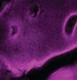
SRS detects molecules by reflecting light waves characteristic of their chemical bonds. The microscope illuminates the test target using two independent laser devices, tuned in such a way that the difference in frequency between their light waves is comparable to the reflection frequency characteristic for the tested molecules.
Wavelength change occurs when a chemical bond in a molecule scatters the light used to illuminate the test object, without the use of luminous markers characteristic of other types of microscopy. While these markers are very versatile and allow specific imaging, unlike SRS where it is impossible to distinguish one protein from another, they often interact with the normal functions and movement of biological molecules. With the SRS, this problem disappears.
However, the use of SRS to date has been limited by the slow imaging rate. The speed of 1 image in 45 seconds was too slow to show a live animal, says Christian Freudiger, a scientist working in Xie’s lab and the first co-author of the study. Even the immobilized animal was too fast to get a clear picture with this technique.
The researchers at Xie’s lab decided to take matters into their own hands. Graduate of the university, Brian Saar currently working at Lincoln Laboratory belonging to the Massachusetts Institute of Technology together with Freudiger built a new amplifier that allows to increase the speed of imaging to 25 frames per second
The team upgraded the light wave detector to better detect scattered light, allowing the microscope to take images from thicker samples. As Freudiger said, before that, it was impossible to see an image while it was being made if the sample was too thick.
This modernization is astonishing, said biomedical engineer Andy Downes from the University of Edinburgh in the UK, who also works on similar microscopy techniques. Downes noted that the new microscope detects 10 times more scattered light than the previous generation SRS presented by Xie’s lab two years ago. This increase in sensitivity will allow the use of a technique in which it will be possible to monitor how the drug penetrates tissues, including cancerous tumors. Downes hopes this discovery will allow drug testing to be carried out without tracers.
Now, Freudiger said, the team is working with bioengineer Eric Seibel of the University of Washington in Seattle to minimize the miniaturization of the entire fiber optic system. For now, a centimeter wide prototype has been created.
Christian Freudiger is currently talking to neurosurgeons about testing the microscope in brain surgery to test its usefulness. We hope that the entire system will be reduced to a width of millimeters – he said. (PAP)