Contents
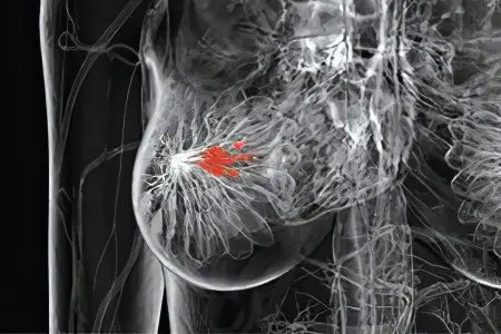
Fibroma is a benign neoplasm that develops in connective tissues. The place of its appearance can be the uterus, ovaries, skin, mammary gland, tendons. In the mammary gland, this tumor manifests itself in the form of a painless lump, having the shape of a ball. In some cases, fibroma is accompanied by a feeling of fullness of the mammary gland on the eve of menstruation. Having found seals in the chest, you should consult a doctor for advice in order to exclude malignant tumors.
Types of breast fibroma

Fibroma formation in the mammary gland is divided into two types: diffuse fibroadenomatosis and fibroadenoma. The first type is characterized by the growth of the tumor throughout the volume of the breast with the presence of compaction and the presence of pain in the mammary gland. The second has a local form of manifestation of fibroma. This is a round formation with a dense structure; when probing, you can feel a movable knot not associated with the skin. The size of this fibroma reaches a diameter of 70 mm.
Fibroadenoma is further classified into several types based on morphological features. The most common types include: pericanalicular, intracanalicular, mixed fibroadenoma, which combines the features of the first two types. The above tumors are characterized by tissue growth around the ducts and the absence of a tendency to degenerate into a malignant formation. The rarest type of fibroadenoma is phylloid, which is characterized by rapid growth and the ability to degenerate into a sarcoma.
Causes of breast fibroma
At the moment, there are no clearly identified causes that form breast fibroids. Numerous observations can only state that the development of the tumor occurs against the background of hormonal failure in the female body. It is at this point that the amount of estrogen in the blood rises. Therefore, girls who are in puberty and women during menopause should be especially attentive to themselves and regularly visit a mammologist.
Diagnostics
There are several stages for diagnosing the disease. First of all, a clinical examination of the woman is carried out. Then, with the help of palpation, a neoplasm is detected. Conducting an ultrasound of the breast gives a detailed idea of the location of the tumor, its exact shape and size, as well as its structure. Next, a biopsy of the tumor is prescribed, which is controlled by ultrasound and is performed using a thin needle using the aspiration method. The resulting material undergoes a cytological examination for the presence of malignant cells.
The diagnosis may require clarification in order to completely exclude diseases such as cysts, cancer, and cystadenopapilloma. In such cases, a core-needle biopsy, X-ray mammography is prescribed, or a diagnostic resection of the mammary gland is performed, followed by a histological examination. It is possible to finally exclude the presence of a cancerous tumor after its removal and laboratory study.
Breast fibroma treatment methods
The method of treatment is determined only after a comprehensive diagnosis based on the results of a morphological conclusion. If a breast fibroma of a small size up to 8 mm is detected, medical treatment controlled by ultrasound is recommended. Its duration can be 4 months or more. However, statistics show that strict adherence to prescribed therapy rarely gives a positive result. Most fibromas are treated with surgery.
The indication for surgery is: rapid growth of the tumor (a twofold increase in fibroma in three months), the size of the formation is more than two centimeters, a cosmetic defect from the presence of fibroma. The reason for the surgical intervention is the leaf-shaped structure of the fibroma, the planned pregnancy (an increase in the tumor can block the milk ducts, the child will be left without feeding, the milk will stagnate, which threatens to develop cancer), the woman’s desire to get rid of the fibroma.
Today, there are two methods of tumor removal. The first is a sectoral resection, which is performed if there is a suspicion of a cancerous tumor. When it is carried out, not only the fibroma is removed, but also the surrounding tissues. The second – enucleation of the tumor, is carried out in the complete absence of suspicion of a malignant tumor. Enucleation takes place under local anesthesia. When it is carried out, only the fibroma is removed, and the postoperative period does not require long-term treatment in a hospital. After such an operation, traces of sutures are not visually noticeable.
Prevention
There are no clear methods of prevention, but if each woman independently palpates the breast or regularly visits a mammologist for a routine examination, this will significantly reduce the risk of developing fibroids and help avoid serious complications.


