Contents
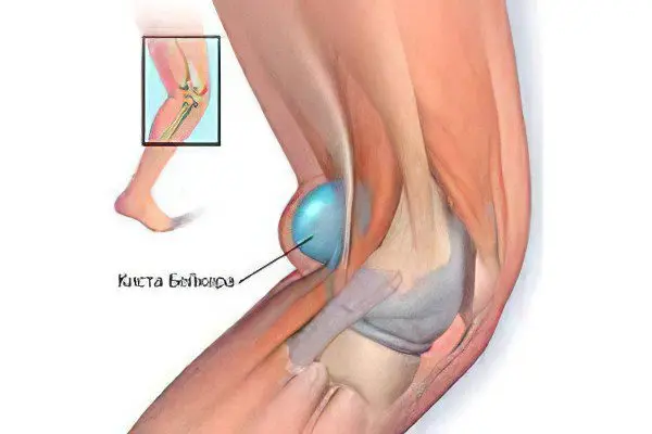
A cyst of the knee joint is a tumor-like benign formation that is localized on the back wall of the joint and is an accumulation of joint fluid. Clinically, this phenomenon is characterized by swelling of the popliteal fossa. Since the cyst has a connection with the articular cavity, its protrusion is in many ways similar to a hernia. The knee cyst can range in size from 2 to 100 mm. Reaching a large size, the formation bursts.
The knee cyst is most clearly visualized when the leg is extended; it is more difficult to detect it at the moment of flexion. The skin in the place of localization of the formation does not change its color, there are no adhesions.
Suffer from this disease mainly those people whose work activity is associated with high physical activity, carrying heavy loads, as well as athletes. In children, this disease is much less common than in older people.
A knee cyst can be a secondary disease that develops against the background of such diagnoses as arthrosis, arthritis, and the like.
A cyst of this kind is very labile: it has the ability to change size or disappear. Cystic formations themselves are single (when only one cavity is formed) and multiple (several small cysts appear).
Signs and symptoms of a knee cyst

This disease has the following manifestations:
swelling of the joint area with clear boundaries, which lends itself well to palpation;
impaired functioning of the joint of varying degrees, up to immobility;
soreness during the work of the joint;
swelling and numbness of nearby tissues.
In this case, symptoms may be completely absent at the initial stage or manifested due to mild discomfort. But with an increase in the size of the cyst, pressure on the nearby nerves and vessels increases, which leads to numbness, tingling in the sole, pain in the knee itself, and a feeling of constant cold in the area below the knee. Movement of the leg becomes difficult and causes pain.
Occasionally, the pressure of the knee cyst on the popliteal vein is so intense that it contributes to the occurrence of deep vein thrombosis or varicose veins. This situation is accompanied by a feeling of discomfort, heaviness, swelling.
With such a complication as a rupture of a cyst of the knee joint, severe, sharp pain can occur, the skin on the back of the leg turns red, swells, and the temperature rises.
Causes of a knee cyst
The following factors that are associated with the processes of the pathology of the knee joint can cause the occurrence of a cyst:
injury to the knee joint: blows, subluxations, which are most often associated with playing sports;
degenerative changes and damage to the menisci;
chronic stage of inflammatory processes in the membranes of the knee joints;
damage, destruction of the cartilage of the joint;
the presence of diseases such as rheumatoid arthritis, osteoarthritis, patellofemoral arthrosis, etc.
Due to these circumstances, increased production of synovial fluid begins, and its accumulation in the back of the knee. A large amount of this substance begins to put pressure on the endings of the nerves, which leads to the appearance of characteristic symptoms.
Excess fluid in the knee joint is also called joint effusion or water in the knee.
A knee cyst can occur even when the knee is generally healthy. At the same time, the bursa – sacs of synovial fluid designed to reduce friction of the moving elements of the joints – forms a tunnel that connects to the joint membrane. Through it, the fluid flows into the knee bursa, causing swelling. This clinical picture is most typical for pediatric patients.
To identify the exact cause of the disease allows ultrasound of the knee joint or MRI. Less commonly, for this purpose, a puncture of the contents of the cyst is performed.
Baker’s cyst of the knee
A Baker’s cyst is a soft but hard to the touch benign lesion that develops in the popliteal fossa (back surface of the knee joint). It is characterized by an elastic dense structure and is characterized by deformation of the synovial membrane and destructive processes in the joint capsule.
Baker’s cyst resembles a hernia in structure, since it does not consist of individual cells, but has a connection with the articular bag.
This formation is best seen when the knee is extended, when it is bent, the cyst is less bulging and has a lower density.
This disease was named after the British physician William Baker, who owns the first publications with reports on synovial cysts localized in the knee joint.
Other types of cysts of the knee joint

Ganglion cyst of the knee It is considered a benign formation that comes from the tendon sheath and joint capsule. It is a spherical or oval formation with a duct in the middle that connects the cyst to the tendon sheath and articular capsule. The cyst is filled with a clear liquid or gelatinous substance. When examined, a ganglion cyst looks like a small water sac or a tight elastic tumor. This disease is quite rare. It can be difficult to determine its cause unequivocally, most often young women suffering from joint hypermobility are susceptible to it. In addition, a ganglion cyst of the knee joint is sometimes the result of an injury.
Parameniscus knee cyst occurs when the meniscus cyst extends to the ligaments and capsular zone. The tumor-like formation becomes large and does not disappear when the knee is extended. Palpation and diagnosis are not difficult.
Parameniscal cyst of the knee joint is referred to as the third stage of cystic degeneration of the meniscus. A complex form of the disease requires surgical intervention. It can be avoided with timely access to a doctor, correct diagnosis, an integrated approach to treatment with subsequent physiotherapeutic measures. In this case, it is possible to achieve a complete restoration of the functions of the knee.
Synovial cyst of the knee may be characterized by a hernia or a change in the synovial membrane of the joint. The exact cause of this disease is not clear, but people suffering from osteoarthritis, rheumatoid arthritis and joint injuries are at risk. Since conservative methods of treatment give a weak effect, most often they resort to operational measures. During the operation, the synovial cyst is removed and the weak point of the capsule is sutured with a special suture that strengthens it.
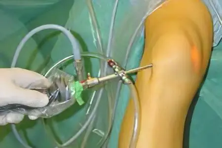
Modern medicine offers endoscopic removal of the synovial knee cyst, since this method allows minimizing injuries and speeding up and improving the efficiency of the rehabilitation process.
It is important to seek medical help in time, since a synovial cyst can rupture at any time and thereby significantly complicate the treatment and restoration of knee functions.
Ankylosing spondylitis in the knee joint is a rarer name for a Baker’s cyst. This is inflammation of the articular cavity caused by various dystrophic processes with the formation of a tumor.
Subchondral knee cyst recognized as a benign cystic lesion, characterized by multiplicity. This formation consists of fibrous tissue filled with myxoid content or silicone. It accompanies the course of a number of degenerative changes in the articular cartilage.
A subchondral cyst is more common in older people and most often has an asymptomatic course. The diameter of the formation can vary from 0,5 to 1,5 cm. During the examination, a defect in the subchondral bone is observed, for which the walls are the surrounding normal bone tissues. The outer fibrous tissue has a loose or dense texture, and the inner lining of the subchondral cyst is not defined.
Cyst of the medial meniscus of the knee joint reveals itself as pain, localized in the inner region of the knee joint, which is felt especially acutely with intensive flexion of the leg. A symptom may be weakness of the anterior thigh muscles. Such a cyst usually protrudes in front of or behind the inner side wall. The most effective treatment is arthroscopy, which provides a small incision size and speed of recovery without a plaster cast.
Treatment of cysts of the knee joint
If there are symptoms that indicate the appearance of benign formations, you should immediately seek help from competent specialists. Since the treatment of a cyst is always selected individually, taking into account the characteristics of a particular case, only a qualified doctor can choose the most suitable from the list of effective methods. This may be conservative treatment or surgical measures.
In most cases, drug treatment is ineffective. Various drugs can help reduce pain, but they do not affect the cause of the disease, which means they are not able to restore mobility to the joint. This method is used mainly before or after surgery. Then prescribe drugs that relieve inflammation and relieve pain.
One of the surgical methods is cyst puncture. It is carried out as follows: a cyst is pierced with a thick needle and the fluid is sucked out of it. Then steroid anti-inflammatory drugs are injected into the intertendon bag. A puncture can only give a temporary result, since in the future the load on the knee joint will lead to the fact that the cavity will again be filled with joint fluid.
Physiotherapy is prescribed only during the period of remission, since physical activity can cause the resumption of pain and complications of the disease.
Therapeutic exercises for a cyst of the knee joint are prescribed by a physiotherapist and at first monitors their implementation. Later, the patient can do exercise therapy at home and conduct light self-massage sessions.
In order to reduce pain and relieve inflammation, UV radiation, infrared laser therapy, and low-intensity UHF therapy are used. Hydrogen sulfide, radon baths are prescribed to improve blood flow.
Removal of a cyst of the knee joint

Indications for surgical intervention and removal of the cyst are the following cases:
education is large;
the cyst is very painful;
the disease progresses rapidly;
other methods of treatment were ineffective.
During the operation, the cyst is completely removed, local anesthesia acts. The duration of the operation reaches half an hour, the next day the patient leaves the hospital, after 5 days the knee joint is ready for minor loads, after a week the sutures are removed.
Removal of a knee cyst may be recommended as a treatment for an underlying knee problem, such as a torn meniscus, as the formation is a secondary disease in this case.


Paldies , ļoti informatīvā publikācija!