Contents
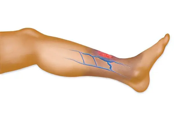
Thrombosis of the sural vein is a blockage of its lumen by a blood clot that disrupts the normal flow of blood. The sural veins are a separate group of deep veins that are located in the thickness of the soleus and gastrocnemius muscles. The diameter of the veins is quite impressive and can reach 10 mm. Venous walls are thin. The sural veins have a close relationship not only with the rest of the intramuscular veins of the lower limb, but also with the superficial veins. Thrombosis of the sural veins is important in the pathogenesis of chronic venous insufficiency of the lower extremities. In the sural veins, embologenic thrombi can be located, which often do not give symptoms, but pose a threat not only to health, but also to the life of the patient.
In the practice of modern clinicians, thrombosis of the sural veins is quite common, especially in comparison with thrombosis of the upper half of the body. The pathological condition is expressed by severe pain, blue skin, an increase in the size of the superficial calf veins. If such symptoms occur, it is necessary to seek immediate medical help in order to receive adequate therapy for the disease.
Causes of sural vein thrombosis
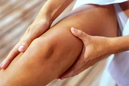
In order for a person to develop thrombosis of the sural veins, a combination of several pathogenetic factors is necessary:
Damage to the inner layer of the vein. High blood pressure, penetration of bacterial endotoxins into the blood, exposure to radiation, metabolic disorders, toxic effects of cigarette smoke, etc., can lead to such injuries.
Violation of blood flow. Blood stasis is one of the most significant causes that lead to the formation of blood clots in the veins. Blood turbulence, myocardial infarction, rheumatic mitral valve stenosis, polycythemia vera, sickle cell anemia have a negative effect on the veins.
Hypercoagulability of blood (thrombophilia). Causes of blood hypercoagulation: high levels of fibrinogen in the body, prothrombin mutations, impaired fibrinolysis, etc.
There is a high probability of developing thrombosis in people who are forced to adhere to bed rest for a long time period. This is especially true for hospital patients. However, even a long flight in an airplane can have an effect. Doctors call deep vein thrombosis an “economy class disease” for a reason. Therefore, any long pastime in a sitting position with legs down can result in thrombosis of the sural veins.
Risk Factors
Other factors that cause a high risk of thrombosis of the sural veins:
Postponed myocardial infarction;
Atrial fibrillation;
Damage to the tissues of the lower extremities, including deep burns, leg fractures, surgical interventions on the veins and soft tissues;
The presence of a malignant tumor in the body;
Presence of a prosthetic heart valve;
Cardiomyopathy;
Nephrotic syndrome;
Elevated levels of estrogen in the blood during pregnancy and in the early period after childbirth;
Burger’s disease;
Obesity and diabetes;
Frequent infectious diseases;
Excessive physical activity and hypodynamia;
Taking hormonal drugs to prevent unwanted pregnancy;
Smoking;
Elderly age.
Symptoms of sural vein thrombosis
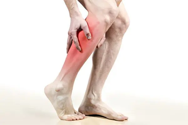
Thrombosis of the sural veins is expressed by the following symptoms:
Pain localized in the limb, the veins of which are affected by a thrombus. The pain is bursting in nature, occurs in the calf area.
If you try to put pressure on the leg along the vein, the pain will intensify.
Pain increases during movement.
The calf muscle may become hot to the touch. Sometimes a person suffers not only from local hyperthermia, but also from a general increase in body temperature.
Swelling in the calf muscle. Moreover, the leg can begin to increase in size literally before our eyes, if we are talking about acute thrombosis of the sural veins.
Enlargement of superficial veins in size.
It should be taken into account that thrombosis of the sural veins is not always accompanied by all of the above symptoms. Moreover, in 50% of patients, blood finds an additional exit into the saphenous veins through the system of communicating veins. This allows you to partially normalize blood flow bypassing the affected vein and minimize the symptoms of pathology. Therefore, the fact that a person has thrombosis of the sural veins can only be recognized by the developed vascular branches of the blood vessels translucent through the skin. They will be located in the lower leg and calf muscles.
Why is sural vein thrombosis dangerous?
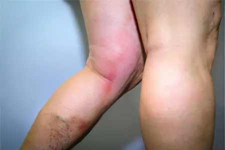
Chronic venous insufficiency of the lower extremities is the most common complication of sural vein thrombosis. This condition leads to the appearance of pronounced edema, malnutrition of the tissues of the lower extremities. As a result, the patient’s legs may develop eczema, non-healing trophic ulcers for a long time. In addition, a person will constantly experience heaviness in the legs, at night he will be disturbed by convulsions. The skin will lose its elasticity, become dry, covered with pigmented rashes. This will lead to the fact that the patient will hardly endure both physical and mental stress.
An even more formidable complication of sural vein thrombosis is pulmonary embolism. In this case, pieces of a blood clot break off, which rise higher and higher through the bloodstream, reach the pulmonary artery and block its lumen. As a result, the patient develops pulmonary and heart failure, which leads to death. If a particle of a blood clot clogs a small branch of the pulmonary artery, then a person develops such a serious condition as a pulmonary infarction.
Diagnostics
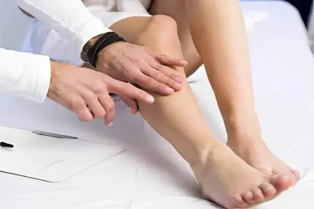
To diagnose thrombosis of the sural veins, the doctor examines the patient.
Specific methods that can be carried out directly at the time of admission are the following:
Positive Homans sign. It is characterized by pain that occurs in the calf muscle during flexion of the foot. The patient is in the supine position.
A positive Moses test, when the patient feels pain when squeezing the lower leg in the direction from front to back. If you press on the shin in the lateral direction, pain will not occur.
Other research methods: Lowenberg’s test, Lisker’s sign, Louvel’s sign, marching test, Pratt’s test – 1, Mayo-Pratt’s test.
These examination methods allow the specialist to suspect deep vein thrombosis of the lower extremities.
To clarify the localization of the thrombus, instrumental diagnostics will be required:
Phlebography.
duplex scanning.
Ultrasound of the veins of the lower extremities.
Radionuclide scanning.
Rheovasography of the lower extremities.
As a rule, doctors prefer phlebography, which allows you to clarify the degree of damage and the location of the thrombus.
Treatment of sural vein thrombosis
If thrombosis of the sural veins has an uncomplicated course, then treatment is possible exclusively by conservative methods. But at the same time, the patient must observe strict bed rest for two weeks, but no less.
For therapy, the following drugs are used:
Direct anticoagulants. They contribute to a decrease in blood viscosity, as well as increased release of antithrombin in the body. As a result, the thrombus dissolves. They are administered in the form of droppers in a hospital setting.
Indirect anticoagulants. These are milder anticoagulants, prescribed in the form of tablets. They prevent the production of thrombin. It should be taken into account that their intake is associated with a risk of bleeding, so the patient must be under strict medical supervision.
Only in a hospital setting can a doctor prescribe streptokinase and urokinase (administered only in the form of injections). They allow the blood clot to dissolve and prevent blood clotting.
NSAIDs are drugs that aim to reduce inflammation, thin the blood, and reduce pain.
Thrombolytic drugs can be prescribed only in the early stages of thrombus formation. If time is lost, then taking them is associated with the risk of a blood clot breaking off. An elastic bandage is applied to the affected limb and lifted. Compression stockings can serve as a replacement for bandages.
Operative therapy
Sometimes, with thrombosis of the sural veins, it is not possible to avoid surgical intervention. The help of a surgeon is required in the following cases:
The patient develops thrombophlebitis.
There is a high risk of developing pulmonary thromboembolism.
The presence of a floating thrombus. This means that it is not fixed on the vascular wall, so it can come off at any time.
The circulation of the limb is severely impaired.
The operation to remove a blood clot is called a thrombectomy. It is contraindicated in the case when the patient has decompensated diseases of the cardiovascular system or respiratory system, or an acute infection is observed.
After discharge from the hospital, the patient must strictly comply with all medical recommendations, as well as eat right, enriching his diet with fortified foods. Seafood must be present in the diet, as they contain copper, which is needed by the vessels to maintain their elasticity. Be sure to refrain from smoking and drinking alcohol.
[Video] Cardiovascular surgeon, phlebologist Abasov M. M. – Products that thin blood clots:


