


The skin is the largest organ in the human body. A physical examination without examining it closely is incomplete. Therefore, it is not surprising that in many diseases of internal organs, changes appear on the skin, the exact knowledge of which often makes it easier to make a correct diagnosis.
SKIN CHANGES IN INTESTINAL DISEASES
Inflammatory bowel diseases
It is estimated that approximately 5,2% of patients with inflammatory bowel disease (Crohn’s disease, ulcerative colitis) show mucosal lesions, and 11% have skin lesions. They can either precede, occur simultaneously with the onset of the symptoms of the underlying disease, and develop later in it. The changes in the skin and mucous membranes accompanying intestinal diseases can be divided into four groups:
1.Diseases for which the etiology of Crohn’s disease / ulcerative colitis is not fully understood (appears to represent some form of allergic reaction to unknown allergens associated with inflammatory bowel disease) resulting from reduced nutrient absorption;
2. secondary to the treatment applied;
3. skin lesions that are an integral part of Crohn’s disease;
4. disorders whose etiological relationship with Crohn’s disease / ulcerative colitis is not fully understood.
- Make an appointment with a dermatologist today! Get free advice
Nodular erythema (see chapters V and XVI)
Syn.: Erythema nodosum.
Def .: Inflammation of the subcutaneous tissue in the form of painful, vivid red or bluish tumors, most often located on the anterior surface of the lower leg.
Epid .: Erythema nodosum is the most common cutaneous manifestation of inflammatory bowel disease. It occurs in about 10% of patients with this disease, more often in women. Skin lesions sometimes precede the diagnosis of intestinal disease (rarely – gastrointestinal symptoms), and their severity does not reflect the severity of the underlying disease.
Erythema nodosum can be caused by:
a. bacterial (mainly streptococcal, Yersinia enterocolitica, tuberculosis) and fungal infections (e.g. histoplasmosis);
b. inflammatory bowel diseases;
c. use of certain medications (most often contraceptives and estrogens, also sulfonamides, salicylates);
d. sarcoidosis (Löfgren’s syndrome is a type of sarcoidosis in which erythema nodosum and bilateral hilar lymphadenopathy, as well as fever, cough and joint pain) coexist.
Etiol .: Allergic reaction to various triggers that vary with population and geography. In 60% of cases, it is impossible to identify them.
Localization: The typical location of the lesions is the anterior surface of the lower leg (usually symmetrically). However, tumors can also occur on the trunk, forearms and face.
Clinical: Primary lesions are tender, not clearly demarcated tumors. Initially vivid red, then they turn brown, and when they give way, they turn yellow-green. They are usually multiple, a few centimeters in diameter, with no tendency to decay and ulcerate. They usually last for 2 to 6 weeks. They resolve without scarring, and there may be post-inflammatory discoloration. The disease may be accompanied by increased body temperature and joint pain.
Hist .: Septic cellulitis is present. Early infiltration includes lymphocytes, eosinophils and neutrophils. Sometimes small granulomas can be found.
DL .: There are no characteristic abnormalities in laboratory tests. The most frequently performed tests: complete blood count, red blood cell decontamination – ESR (usually elevated), antistreptolysin test – ASO, determination of liver enzymes, throat swabs, stool cultures, tuberculin test, chest X-ray, colonoscopy.
DR .: Other types of cellulitis, nodular vasculitis, erysipelas, erythema induration.
Treatment: Topical: topical glucocorticosteroids, compresses with 2% ichthyol solution, ichthyol ointment. Supportive: bed rest, limb elevation, compression stockings. General (depending on the causative factors): non-steroidal anti-inflammatory drugs (aspirin, naproxen, indomethacin), supersaturated potassium iodide (5-10 drops 2 times a day), general corticosteroids (usually prednisone at an initial dose of 40 mg daily), broad-spectrum antibiotics .
Year: Mileage is usually self-limiting. Occasionally there are relapses.
Pyoderma gangrenosum (Fig. 31.1)
Syn.: Pyoderma gangrenosum.
Def .: Chronic progressive skin necrosis often associated with the underlying disease.
Epid .: Pyoderma gangrenosum occurs in 0,5-20% of people with inflammatory bowel diseases (most often ulcerative or pustular), slightly more often in women. It usually appears after revealing symptoms of the underlying disease. It is often associated with systemic diseases, such as inflammatory bowel diseases (the most common), other gastrointestinal diseases (hepatitis C, chronic active hepatitis, primary biliary cirrhosis, carcinoid syndrome, gastric and duodenal ulcer), haematological diseases (leukemia, polycythemia vera, lymphomas), arthritis, immunodeficiency.
Etiol .: Unexplained. The role of an excessive inflammatory reaction caused by immune abnormalities has been suggested (the pattern is found in approximately half of the patients).
“FIGURE 31.1. Pyoderma gangrenosum. “
Loc: Most often on the front surfaces of the lower limbs, buttocks or the trunk (but also in the area of postoperative wounds, in the genital area, on the head and neck).
Klin .: There are four clinical forms of pyoderma gangrenosum:
a. ulcerative form – the primary lesion is a red, inflamed lump or pustule that rapidly spreads peripherally and ulcerates. The resulting very painful ulcer is deep, well demarcated from the surroundings, with a necrotic bottom filled with purulent contents and purple-colored undermined edges. Sometimes the changes are numerous. Some may heal leaving parchment-like scars, while others may form new scars. The course of the disease may be electrifying, and in untreated cases, muscles, fascia and even bones may be exposed. In many patients, the changes are accompanied by fever, malaise and joint pain, and their occurrence is preceded by a slight trauma;
b. pustular form – characterized by painful, sterile pustules that do not ulcerate;
c. bullous form – initially a bladder, from which ulceration rapidly develops (often coexisting with malignant hematological growths);
d. rocking form – initially shallow ulcer slowly develops papillary hyperplasia.
Hist .: Visible infiltration of neutrophils within the peripheral part of the lesion and histological signs of vasculitis. In addition, there may be small vessel thrombosis. Later, significant tissue destruction develops with massive infiltration of neutrophils, lymphocytes, plasma cells and giant cells.
DL: Laboratory tests are important in the search for possible underlying diseases: complete blood count, ESR (often accelerated), indicators of liver and kidney function, colonoscopy, skin biopsy (to exclude deep fungal infections) and bone marrow biopsy.
DI .: Immunoglobulins and complement components are found in the vessel walls from the edge of the wounds.
DR .: Vasculitis, Sweet’s syndrome, bacterial infections, deep mycoses, panniculitis.
Healing: Topical: steroids, tacrolimus. Supportive: limb elevation, rest. Surgical interventions should be avoided, as they may cause enlargement of the lesions.
General: corticosteroids administered orally (usually prednisone at an initial dose of 60-80 mg / day) or in the form of pulses, dapsone, sulfasalazine (also in patients without inflammatory bowel disease), cyclosporine, IV immunoglobulins
Year: Effective treatment of accompanying illnesses may lead to regression of the lesions. Occasionally, however, there may be chronic relapses that are difficult to treat.
Sweet’s team
Syn .: Dermatosis acuta febrilis neutrophilica, acute febrile neutrophilic dermatosis.
Def .: Acute febrile illness characterized by the presence of intensely red, oedematous-infiltrative lesions on the skin.
Epid .: Most of the patients are women between the ages of 30 and 50.
Sweet’s syndrome can be caused by:
a. inflammatory / autoimmune diseases (Crohn’s disease, ulcerative colitis, cirrhosis, subacute thyroiditis);
b. infections (caused by staphylococci, streptococci, Yersinia, Salmonella, HIV, CMV);
c. malignant neoplastic processes (acute / chronic myeloid / lymphocytic leukemia, lymphomas, breast cancer, ovarian cancer, prostate cancer, thyroid cancer);
d. other causes (drugs, pregnancy, vaccinations).
Etiol .: Sweet’s syndrome is believed to be the result of the immune system being hypersensitive to various factors. GM-CSF (activator of the differentiation of stem cells to mature neutrophils) and IL-1 play an important role in the pathogenesis. A link with the HLA-B54 antigen also seems likely.
Localization: The lesions are most often located on the trunk, upper limbs (on the extension side), as well as the face and neck.
Clinical: Skin lesions appear rapidly and have the form of tender, sharply demarcated, intensely red edematous-infiltrative foci, usually multiple, with a diameter of 4 mm to over 12 cm. The surface of the lesions may peel off, and hemorrhagic vesicles and pimples often form around the periphery. Usually there are general symptoms: fever, muscle and joint pain, nausea and a feeling of general discomfort. The kidneys, eyes, lungs and liver can also be affected.
Hist .: Intense neutrophil infiltration within the skin.
DL: Infections and neoplastic processes should be ruled out. The results of laboratory tests are not specific, but it is recommended to perform a complete blood count with a smear (leukocytosis of the order of 20 / mm000 with a predominance of divided neutrophils, often above 3%), ESR, renal and hepatic function indicators, urine tests (proteinuria and hematuria).
DI .: Deposition of immunoglobulins and complement components in the vascular walls.
DR .: Drug reactions, cellulitis, pyoderma gangrenosum, erythema multiforme, vasculitis, skin leukemia.
Treatment: General: oral corticosteroid therapy (usually prednisone at an initial dose of 40-80 mg / day) or much less frequently: dapsone, cyclosporine, methotrexate.
Year .: Depends on the severity of the accompanying systemic disease. Most patients recover quickly from oral steroids, and may persist for 6-8 weeks if left untreated. Relapses are observed in 25-50% of patients.
Acquired blistering epidermis separation
Syn.: Epidermolysis bullosa acquisita (EBA).
Def .: Chronic, scarring bladder disease in which lesions are mainly located in areas exposed to mechanical injuries.
Epid .: It occurs in adults, slightly more often in women.
Etiol .: An association with the HLA-DR2 antigen is observed in some patients. EBA may be associated with the occurrence of other diseases: Crohn’s disease, diabetes, systemic lupus erythematosus and others.
Loc .: Areas exposed to mechanical injuries: elbows, knees, hands and feet. In a large proportion of cases, the oral mucosa is also affected.
Clinical: Skin lesions have the character of large and tight blisters which, while healing, leave atrophic scars and milia. They are usually accompanied by itchy skin. The general condition of patients is usually good.
Hist .: Subepidermal blisters, inflammation of various degrees.
DI .: Antibodies (mainly IgG class) directed against collagen VII, present in the skin and usually also in the serum of sick people. In the skin split method, they react with the bottom of the artificially created bladder.
DR .: Pemphigoid, scarring pemphigoid, linear IgA dermatosis, pemphigus vulgaris.
Healing: Oral glucocorticosteroids (prednisone), cyclosporine A, dapsone, colchicine, vitamin E, intravenous infusions of immunoglobulin G.
Year: The disease is chronic and difficult to treat. In the case of coexistence of systemic diseases, the prognosis depends on their type.
Dermatitis herpetiformis (see chapter XIV)
Syn .: Duhring’s disease, painful polymorphic dermatitis.
Def .: Chronic multiform dermatitis accompanied by gluten-dependent enteropathy, generally clinically asymptomatic.
Epid .: The disease usually begins in young adults. The male to female ratio is 3: 2. Sometimes the disease is associated with lymphomas of the gastrointestinal tract, inflammatory bowel disease, or autoimmune diseases (e.g., thyroid gland, diabetes, lupus erythematosus, or vitiligo).
Etiol .: The role of genetic factors (about 80% of patients have HLA-B8 and DR3 antigens, which also predispose to the development of celiac disease) and environmental factors (skin changes caused by gluten).
Loc .: The lesions are located symmetrically on the elbows, knees, upper back, buttocks, and on the scalp at the hairline and on the back of the nape.
Clinical: Lesions are polymorphic. There are papules, erythema, urticarial eruptions and small vesicles, arranged in festoons and herpes. Sometimes large blisters form. Skin eruptions are accompanied by intense itching, so sometimes the only lesions are foci covered with scabs. Skin changes often worsen after consuming products containing gluten or potassium iodide. Most patients have gastrointestinal abnormalities similar to those found in celiac disease – celiac disease (villi atrophy of the small intestine), usually asymptomatic.
Hist .: Microrysts in the dermal papillae. Blood vessels surrounded by lymphohistiocytic infiltrates with an admixture of neutrophils and eosinophils. The presence of sub-epidermal blisters.
DI .: In direct immunofluorescence examination, granular IgA deposits are visible (even in remission) at the tips of the dermal papilla (they are also detected in unchanged skin). In addition, the serum shows IgA autoantibodies to endomysium (EMA), tissue transglutaminase (tTG) and gliadin (these antibodies are also present in celiac disease), and often IgA and IgG anti-reticulin antibodies.
DR .: Scabies, eczema, lichen dandruff, erythema multiforme, bullous drug reactions, bullous pemphigoid, pemphigus vulgaris.
Treat: Topically: steroid preparations. Overall: gluten-free diet, dapsone (100-150 mg / day) or sulfapyridine.
Year: The disease persists throughout life with varying degrees of severity. Careful control of patients is recommended due to the risk of gastrointestinal lymphomas and autoimmune diseases.
Aphthae (Fig. 31.2)
Syn.: Aphthae, aphthous ulcers.
Def .: Painful, recurrent mouth ulcers.
Epid .: They occur in approximately 4,3% of patients with inflammatory bowel disease. Aphthas may occur in the course of the following disease states: gastrointestinal diseases (Crohn’s disease, ulcerative colitis, celiac disease); hematological diseases (pernicious anemia, iron deficiency, folic acid deficiency); immunodeficiency (HIV, immunosuppressive therapy); Behçet’s syndrome.
Etiol .: Unknown. Cellular immunity disorders and injuries of the oral mucosa (e.g. cheek biting) are likely to be involved.
“FIGURE 31.2. Tongue canker “
Loc .: The oral mucosa: lips and cheeks, soft palate, tip of the tongue, frenulum, floor of the mouth, tonsils.
Clinical: The primary lesion is a red spot, from which a tiny follicle develops. After its resolution, ulceration appears, 2-5 mm in diameter, round or oval in shape, with a clearly marked erythematous and raised edge. The ulcer is covered with a yellow fibrous substance. The mouth ulcers are usually single or sparse. They disappear after 1-2 weeks without leaving any scars. In addition to the above-described small canker sores, we also distinguish: large (Sutton’s canker sores), which are larger, deeper, very painful, sometimes multiple and disappear with scars, and herpes, which occur in clusters.
DR .: Viral infections (herpes simplex, cytomegalovirus or Coxsackie); chronic ulcerative stomatitis; erosive lichen planus.
Healing: Topical: drying agents, astringents, antibiotic solutions (e.g. 5% tetracycline solution), topical glucocorticosteroids.
Year: Afts are a chronic and recurrent condition that can sometimes make eating, drinking and speaking difficult.
Pyostomatitis vegetans
Def .: Rare, chronic, purulent disease of the oral mucosa associated with inflammatory bowel diseases. It is probably a form of pyoderma gangrenosum.
Epid .: It occurs in people of all ages, slightly more often in women. The lesions usually precede the diagnosis of IBD. It may accompany Crohn’s disease or ulcerative colitis.
Etiol .: Unknown.
Localization: Most often the mucosa of the lips, gums and cheeks.
Clinical: Within the red, swollen and deeply folded oral mucosa there are numerous small spots that may form ulcers.
Hist .: Intra-epidermal pustules (rich in eosinophils) and pseudoplastic epidermal hyperplasia.
DL .: Peripheral blood count (often transient eosinophilia), colonoscopy.
DR .: Afty, pemphigus vulgaris, pemphigoid.
Heal: Necessary treatment of underlying disease. In the absence of intestinal symptoms, treatment with high doses of oral steroids is effective.
Less commonly, inflammatory bowel diseases are accompanied by the following conditions:
a.vesicular pustular eruptions,
b. leukocytoclastic vasculitis,
c. polyarteritis nodosa,
d. erythema multiforme,
e. lichen planus.
Skin changes resulting from impaired food absorption
They are associated with the involvement of the small intestine by the disease process and resections of the affected sections of the intestine, which in turn leads to deficiencies of iron (anemia), vitamins (e.g. pellagra), albumin (edema) and zinc.
Zinc deficiency (Fig. 31.3)
Def .: Hereditary or acquired decrease in the level of zinc in the body, which results, among others, in to the development of skin lesions in the distal parts of the limbs and within natural foramen, and diarrhea.
Etiol .: The cause of the disease is zinc deficiency in the diet or its impaired absorption (e.g. in inflammatory bowel diseases, chronic diarrhea, pancreatitis or chronic kidney diseases). An autosomal recessive inheritance of intestinal zinc absorption is known as acrodermatitis enteropathica.
Loc: Area of the body’s natural orifices (around the mouth, nostrils, eyes, anus, genitals) and the distal parts of the limbs (fingers and toes, palms, soles).
„RYC.31.3. Acrodermatitis enteropathica.”
Clinical: The skin lesions are erythematous spots, sharply delimited from the surroundings, covered with erosions and scabs in the central part, and with vesicles and pustules on the periphery. Sometimes there are psoriasis-like lesions. Secondary superinfections with Candida yeasts and staphylococci are often found. Skin appendages may also be involved. Among the nail changes, pustular rot with subsequent dystrophy of the nail plates should be mentioned. The hair becomes fine and thinning, sometimes with light and dark strands (similar to the color of a zebra), reflecting changes in the levels of zinc in the body. Sometimes there is inflammation of the tongue and oral mucosa and conjunctivitis. The most common systemic symptoms include: diarrhea of varying severity, apathy, irritability, impaired wound healing, and stunted development in children.
Hist .: Significant parakeratosis is observed. The epidermis is spongy and acantotic, and its upper layers are necrotic and pale in color. In the papillary layer of the skin, lymphohistiocytic perivascular infiltrates are found.
DL .: A pathognomonic symptom is a low level of zinc in the serum, which also shows a low concentration of alkaline phosphatase (zinc-dependent metalloenzyme).
DR .: Seborrheic dermatitis, psoriasis, Candida infections, biotin deficiency, fatty acid deficiency, migratory necrolytic erythema.
Healing: Administering oral zinc preparations (standard dose is 30-150 mg / day).
Year: With the use of pharmacological zinc supplementation, a satisfactory general condition of patients is achieved. Skin lesions secondary to the applied treatment In the treatment of inflammatory bowel diseases, oral glucocorticosteroids, sulfasalazine and immunosuppressants are the most commonly used. Their intake may be associated with the occurrence of side effects. The skin lesions include: steroid acne, cushingoid body structure, stretch marks, hirsutism, secondary infections, leukocytoclastic vasculitis, polyarteritis nodosa, maculopapular exanthema and erythema multiforme. Skin lesions that are part of the Crohn’s disease picture Crohn’s disease develops fistulas (entero-cutaneous, entero-bladder, entero-vaginal), fissures and perianal abscesses. There may also appear erythematous changes, skin hardening and ulcerations, mainly in the vicinity of fistulas and artificial anus, but also in unchanged skin. In the oral cavity, granulomatous changes (sometimes granulomatous cheilitis occur – the lips are swollen and hard on palpation) and gingival hyperplasia.
Non-inflammatory bowel diseases
Peutz-Jeghers syndrome
Syn.: Hamartomatous intestinal polyposis, periorificial lentiginosis.
Def .: A syndrome in which intestinal polyposis is accompanied by pigmented spots.
Etiol .: Inherited autosomal dominant.
Loc .: Pigmented lesions most often located on the oral mucosa, lips and sometimes on the skin around the mouth, eyes and limbs.
Clinical: There are numerous lentil spots and freckles. While skin lesions may tend to subside, mucosal lesions tend to be more persistent. Pigment spots are accompanied by intestinal polyposis (the jejunum is always involved), and it may lead to bleeding and intussusception. The lifetime risk of neoplastic transformation in the gastrointestinal tract exceeds 50%. There is also an increased risk of developing cancers of the genital organs and the breast.
DR .: Peutz-Jeghers syndrome, LEOPARD syndrome.
But: Skin lesions do not require treatment. Systematic gastroenterological and surgical control is recommended.
Year .: Depends on intestinal lesions.
Gardner’s syndrome
Syn .: Familial adenomatous polyposis, familial polyposis of the large intestine.
Def .: A syndrome characterized by the presence of intestinal polyps and numerous sebaceous cysts and fibromas on the skin.
Etiol .: The disease is inherited autosomal dominantly. A gene whose mutations are responsible for its development was identified – APC (tumor suppressor gene).
Clinical: Probably the earliest symptom of Gardner’s syndrome are pigmented changes in the retina, defined as congenital hypertrophy of the pigmented epidermis. Most patients also have osteomas of the mandible, maxilla and facial bones. In addition, there are: epidermal cysts (sometimes huge), desmoid tumors (most often within scars) and fibromas. Within the digestive tract there are multiple polyps of the colon and rectum, which, if left untreated, always turn into colorectal cancer (polyps and cancers can also occur in the stomach and small intestine). Patients are also at increased risk of developing hepatoblastoma.
DR .: Peutz-Jeghers syndrome, Cowden syndrome, common epidermal cysts.
Healing: Screening for polyps in family members predisposed to developing gut polyposis. Prophylactic colectomy is recommended in adolescents with Gardner’s syndrome.
Year: Bad if colon removal surgery is not performed before cancer occurs.
Paraneoplastic skin syndromes associated with intestinal cancer
Leser-Trélat syndrome (Fig. 31.4)
Def .: Sudden sowing of numerous seborrheic warts as a symptom of an ongoing neoplastic process (most often it accompanies adenocarcinomas, mainly of the gastrointestinal tract).
Etiol .: Some cancers of internal organs, especially adenocarcinomas, are responsible for the secretion of growth factors that stimulate the development of seborrheic warts.
Lock: Torso, face, backs of hands, sometimes also hairy scalp and genital area.
Clinical: Seborrheic warts may have different morphology: from flat-raised, well-demarcated lumps of a color similar to healthy skin, to papillary, hyperkeratotic, brown structures, as if superimposed on the skin, sometimes pedunculated.
“FIGURE 31.4. Leser-Trélat syndrome. Sowing of seborrheic warts on the trunk. “
Hist .: Skin lesions do not tend to malignantly transform. There is a mild proliferation of keratinocyte-like cells in the basal layer of the epidermis, which typically reach the surface of the skin.
DR .: Actinic keratosis.
Heal: The underlying disease.
Year: Depends on the stage of the neoplastic process. Diffuse seborrheic warts, unlike other seborrheic warts, may spontaneously regress or reappear when the underlying tumor is removed or recurs.
Muir-Torre syndrome
Def .: Paraneoplastic syndrome in which tumors of various internal organs are accompanied by sebaceous cysts and keratoacanthomas.
Epid .: It most often accompanies cancers of the gastrointestinal tract, lungs and genitourinary system.
Etiol .: Autosomal dominant disease. Numerous mutations in DNA repair genes are found.
Localization: Skin lesions are usually located on the face, less often on the head and torso.
Clinical: There are multiple sebaceous cysts, neoplasms of the sebaceous glands and keratoacanthomas (these are domed tumors, usually not different in color from the surrounding skin, with a characteristic crater-like depression in the middle, filled with horny masses).
But: Oral retinoids may possibly inhibit the growth of skin cancers.
Year: Depends on the stage of the neoplastic process.
SKIN CHANGES IN DISEASES OF THE PANCREAS
Acute, chronic pancreatitis
Pancreatitis, in addition to the leading symptoms such as epigastric pain, increased levels of pancreatic enzymes, vomiting and nausea, may be accompanied by jaundice, ascites, facial erythema, pale blue discoloration around the navel (Cullen’s sign), bruising-like discoloration on the side surfaces of the abdomen (Gray-Turner symptom) as well as reticular cyanosis and panniculitis.
Reticular cyanosis
Syn.: Livedo racemosa, vasculitis racemosa.
Def .: Persistent bruising of the skin, resembling a net and reflecting vessel damage due to closure or inflammation.
Epid .: A rare disease. Reticular cyanosis can occur in the course of many conditions, the most common of which are:
a.pancreatitis, bacterial endocarditis, rheumatic fever,
b. lupus erythematosus, dermatomyositis, Sneddon’s syndrome, anticardiolipin syndrome,
c. polyarteritis nodosa, atherosclerosis, vasculitis,
d. syphilis, tuberculosis,
e. HIV, AIDS,
f. coagulopathy, cryoglobulinemia.
Etiol .: The changes arise as a result of disturbances in the normal blood distribution, most often due to the closure of the deep vascular plexus of the dermis.
Lock: The buttocks, lower back, upper arms and the arch of the foot are the most commonly involved.
Clinical: Irregular, reddish-blue, mesh-like vascular streaks that turn pale under pressure. Lumps and ulcers are rare. Sometimes patients report severe pain, especially when the lesions are located on the feet.
DL .: There are no specific laboratory tests, diagnosis depends on the underlying disease.
Hist .: Vascular occlusion or inflammatory lesions of a segmental nature accompanied by a thickening of the vascular wall.
DR .: Livedo vasculitis, marbled skin.
But: The only effective treatment is to treat the underlying disease. Sometimes anticoagulants, platelet inhibitors, non-steroidal anti-inflammatory drugs, corticosteroids, dapsone, and azathioprine are used.
Year: Depends on the underlying disease.
Inflammation of the subcutaneous tissue
Syn.: Panniculitis.
Def .: A disease with inflammation in the area of subcutaneous adipose tissue.
Etiol .: The cause is probably the release of lipases by the damaged pancreas or its tumor (in the course of pancreatitis / cancer).
Epid .: May also accompany: other systemic diseases (renal failure, lymphomas, leukemia, sarcoidosis, alpha-1-antitrypsin deficiency); vasculitis (e.g. nodular vasculitis); occur under the influence of physical factors or as idiopathic cellulitis.
Localization: The lesions can be localized all over the body (as opposed to inflammations of the subcutaneous fatty tissue on a different background, when they mainly affect the lower limbs).
Clinical: There are painful subcutaneous nodular eruptions, usually red or bluish in color and measuring 1 to 4 cm. The lumps usually don’t ulcerate. Releasing, they may leave discoloration. General symptoms are common: fever, joint pain, serositis and eosinophilia.
Hist .: Presence of fat cells dissolved by lipolytic enzymes (called ghost cells), necrotic adipose tissue cells surrounded by neutrophils with decayed nuclei. Later, the lesions may become calcified.
DL: Serum amylase and lipase levels should be determined and the picture of the pancreas should be examined.
DR.: Cellulitis.
Heal: Causal.
Year: Depends on the underlying disease.
Pancreatic cancer
The symptoms of pancreatic cancer are similar to those that occur in chronic pancreatitis, hence the differential diagnosis of these two disease entities is very difficult. That is why it seems so important to pay attention to skin changes that may accompany pancreatic tumors.
Wandering necrolytic erythema
Syn .: Erythema necroticans migrans, glucagonomy syndrome.
Def .: Paraneoplastic skin disease associated with a tumor in the pancreas that originates in glucagon-producing cells (pancreatic alpha cells).
Epid .: It is more common in women (the ratio of women to men is 3: 1). In a minority of cases, this type of lesion can also be a symptom of liver cirrhosis, pancreatitis or colorectal cancer.
Etiol .: The presence of glucagonoma is detected in almost all patients with migratory necrolytic erythema. In 80% of cases, the tumor is malignant, but usually slowly growing.
Loc .: Flexion surfaces of the limbs, torso and face.
Clinical: Fatigue and weight loss are usually the first symptoms. Irregular erythematous foci appear on the skin, within which flaccid blisters form. As a result of creeping of the epidermis, vivid red erosions are formed, covered with scabs. The lesions spread circumferentially, creating a unique pattern of intersecting bands, often with scabs and scales present. In addition, there may be glossitis, conjunctivitis, chewing, diffuse alopecia and paronychia.
Hist .: The epidermis shows the presence of necrosis, dyskeratotic cells and acantholysis as well as the formation of submerged pimples containing neutrophils. Older lesions have perivascular lymphocytic infiltrates.
DL .: There is a very high concentration of glucagon in the serum. Diabetes (difficult to control with insulin), anemia, and low levels of zinc, potassium, cholesterol and amino acids in the blood are common.
DR .: Zinc deficiency, Hailey-Hailey disease, pemphigus deciduous, Lyell’s syndrome, pustular psoriasis.
Healing: Surgical removal of the pancreatic tumor followed by cytostatic treatment (most often 5-fl uorouracil). The skin lesions are most effectively affected by topical drying suspensions, often with the addition of antibacterial substances.
Year: If the tumor is completely removed, all symptoms, including necrolytic erythema, resolve very quickly. However, they usually recur, as skin lesions usually appear late, in the period of metastasis.
Wandering superficial thrombophlebitis
Syn.: Thrombophlebitis migrans.
Def .: Recurrent thrombophlebitis in various locations, which is often a symptom of the underlying disease.
Etiol .: Unknown.
Epid .: Most of the patients are young or middle-aged people, usually men. It may accompany: pancreatic, lung or prostate cancer, lymphomas or leukemias, Behçet’s syndrome, diseases associated with deep vein thrombosis.
Loc .: Most often the straight surfaces of the lower limbs.
Clinical: Inflammatory changes are found within various, sometimes interconnected, veins that resemble tethers. Individual blooms disappear within 2-3 weeks, but new ones appear.
DR .: Erythema nodosum, cellulitis.
But: The most important thing is to rule out the underlying disease. In addition, low doses of heparin, non-steroidal anti-inflammatory drugs, sometimes antibiotics are recommended, as well as the use of pressure dressings and the patient’s physical activity.
Year: Depends on the course of the underlying disease. Typically, thrombophlebitis is chronic.
SKIN CHANGES IN LIVER DISEASES
Patients with liver diseases present a wide variety of skin lesions, the mechanism of which their formation is often not fully understood. Many of them are specific to particular disease entities, and some reflect the severity of the liver pathology. Therefore, their knowledge cannot be overestimated when making an appropriate diagnosis and monitoring patients’ response to treatment. Cirrhosis of the liver. Skin changes of a vascular nature
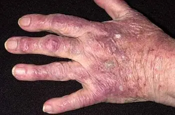
Hands and feet erythema
Syn .: Erythema palmare et plantare symptomaticum, red liver palms.
Def .: Acquired erythema of the hands and feet, which is often a symptom of chronic liver diseases.
Epid .: It can also appear: in pregnant women, in the case of hyperthyroidism, in patients with cancer of internal organs.
Etiol .: Malfunction of arteriovenous anastomoses resulting from disturbed activity of the autonomic nervous system.
Clinical: Palmar erythema (the middle of the hand is usually spared, while the elevations of the withers and the ball, the distal parts of the palms and the tips of the fingers are red) and often erythema of the soles of the feet. Erythematous spots change color according to the rhythm of the heartbeat.
DR .: Familial erythema of hands and feet, erythema of distal regions.
But: Unknown.
Forehead skin erythema
Syn.: Erythema diffusum hepaticum Kalk.
Def .: Acquired erythema.
Epid .: May be observed in some patients with acute liver failure in the pre-coma.
Etiol .: Considered a symptom of hepatic jaundice, the red tinge of which depends on the accompanying dilatation of small vessels.
Clinical: It extends in the space formed by the border of the hair, the bridge of the nose, and the zygomatic bones. It consists of tiny red points. It disappears leaving vascular dilatations and stellate hemangiomas.
Stellate hemangioma
Syn .: Vascular spider, spider nevus, angioma stellatum.
Def .: Acquired vascular lesion characterized by the presence of tiny vessels radiating from the central part.
Epid .: Stellate hemangiomas are observed in about 33% of patients with cirrhosis (in the case of alcoholic cirrhosis in 50% of patients, and in other cirrhosis – in 27%). They can also appear in:
a. 10-15% of healthy children and adults (especially in pregnant women or those taking contraceptives),
b. people with connective tissue diseases, especially with CREST syndrome,
c. patients with hyperthyroidism.
Etiol .: The role of venous blood stasis, hormonal disturbances, substance P and the angiogenic properties of ethanol (in patients with alcoholic cirrhosis) are considered.
Loc .: Face, neck, upper chest and forearms.
Clinical: The stellate hemangioma consists of a slightly elevated pinhead-sized center formed by an arteriole, and small, tapering capillaries radiating from it in all directions. After compressing the lesion with a glass slide, you can see the pulsation of the arteriole. There is often a pale rim around the hemangioma, which is more evident after cooling. Stellar hemangiomas can be single or multiple.
Hist .: We find a dilated central vessel with branches going off.
Treatment: Laser haemangioma removal, electrocautery, and sometimes surgery can be used.
Jellyfish head
Syn.: Medusa’s head.
Def .: Widening of the veins of the abdominal wall.
Epid .: Rare symptom. Widened veins on the inner surface of the abdominal cavity (the so-called inner jellyfish head) are more common (using the color ultrasound method).
Etiol .: Effect of creating a portal-systemic collateral circulation in patients with portal hypertension.
Clinic: Visible radially extending from the navel, dilated and twisted veins under the skin of the abdomen (the so-called outer head of jellyfish).
Heal: The underlying disease.
Hemorrhagic diathesis
Syn.: Purpura haemorrhagica.
Def .: Pathological bleeding tendency.
Etiol .: Toxic damage to the blood vessel wall, increased capillary permeability, deficiency of vitamin K-dependent coagulation factors (II, VII, IX, X), reduction in the number of platelets, levels of prothrombin, phibrinogen and serum albumin.
Clinical: There may be:
a. ecchymosis on the mucous membranes (mouth, nose, conjunctiva) and on the skin (mostly in the lower limbs), disappearing and leaving brown discoloration;
b. bleeding from the gums, nose, gastrointestinal tract and bleeding into the retina;
c. ecchymoses, hematomas and sometimes palpable purple rash. Cases of coexistence of Henoch-Schönlein purpura have also been described.
DL .: Measurement of platelet count, bleeding time, APTT, thrombin and prothrombin time, and phi-brinogen concentration.
DR .: Hemorrhagic blemishes on a different background.
Heal: Treating the underlying disease. Adequate substitution (platelet concentrates, fresh frozen plasma, vitamin K, coagulation factors).
Year .: Depends on the correction of liver function.
Skin color changes
Jaundice
Syn.: Icterus.
Def .: Yellow discoloration of the skin, mucous membranes and sclera due to the deposition of bilirubin in these tissues. The yellowing of the skin becomes visible when the bilirubin level is above 25 mg / dL.
Epid .: May accompany: liver cirrhosis, viral hepatitis, its tumors, toxic liver damage, genetic diseases of the liver (eg Wilson’s disease).
Etiol .: There are several types of jaundice: hemolytic, parenchymal and obstructive. Localization: Skin, mucous membranes and sclera.
Clinical: The degree of yellowish color of the skin varies – from pale yellow or lemon (hemolytic jaundice), through greenish-yellowish (when bile remains in the bile ducts due to mechanical obstruction), to yellowish-red (parenchymal jaundice), and even brown-black (most often in cirrhosis) .
DL .: Determination of serum bilirubin (direct and indirect) and urine bilirubin and urobilinogen.
DR .: Carotenemia, uremia.
But: The most important thing is to remove the cause.
Shame
Syn .: Chloasma, liver spots.
Def .: Limited discoloration of the facial skin.
Epid .: May occur: in chronic liver diseases (not a pathognomonic symptom), in pregnant women, in women using oral contraception.
Etiol .: The dependence of the formation of discoloration on the excess of estrogens in the blood (also influenced by sunlight and genetic factors).
Lock: Temples, forehead, cheeks and upper lip.
Clinical: Initially, small, irregular patterns ranging in color from yellow-brown to brown are visible on the facial skin, which then enlarge and merge into larger clusters. The layout of the changes often resembles a mask.
Hist .: Increased amount of melanin in basal layer keratinocytes and dye incontinence.
DR .: Chronic phototoxic reactions associated with cosmetics, hyperpigmentation due to photosensitivity after certain medications (e.g. phenytoin).
Healing: whitening preparations, sun blockers. Skin changes associated with hormonal disorders
Skin stretch marks (Fig. 31.5)
Syn.: Striae, stretch marks.
Def .: A type of scar with the presence of linear, smooth bands of skin showing signs of atrophy.
Etiol .: Impaired inactivation of steroid hormones within damaged hepatocytes. Other causes of stretch marks: sudden weight gain and associated skin stretching, topical use of strong steroids, symptom of Cushing’s syndrome and Cushing’s disease. It also seems that there is an individual tendency towards their development (especially evident in adolescence).
“FIGURE 31.5. Skin stretch marks on the buttock and right hip “
Lock: Buttocks, tops of thighs, breasts, abdominal wall and skin of the back.
Clinical: The lesions initially appear as streaks of different length and width, red in color, arranged in parallel, which then become white and atrophic. They are slightly sunken and slightly wrinkled. It happens that adipose tissue is emphasized by them.
Hist .: Initially: thinning of the epidermis covering the lesions, mild inflammation, symptoms of elastolysis. Later: collagen fibers in the form of thin eosinophilic bundles running in straight lines.
Healing: Topical preparations (e.g. tretinoin), cosmetic treatment.
Year: The changes are permanent.
Gynecomastia
Syn.: Gynaecomastia hepatica.
Def .: Unilateral or bilateral male mammary gland hyperplasia.
Epid .: It occurs in approximately 10% of men with advanced cirrhosis. Other causes of gynecomastia include:
a.physiological (newborns, puberty, old age, obesity),
b.Symptomatic (prolactinoma, Klinefelter’s syndrome, hypogonadism, hormonally active tumors),
c. drug-dependent (estrogens, tamoxifen, spironolactone, ranitidine, antipsychotics).
Etiol .: Decrease in testosterone, increase in estrogen levels.
Clinical: Breast bulging, enlargement and elongation of the nipples and darkening of the areola are observed. Gynecomastia may be accompanied by: atrophy of the testicles, impotence and hair changes (the hair on the pubic mound may be female – Chvostek symptom, hair in the armpits and on the breasts partially / completely disappears, there is also a complete absence of hair on the abdomen).
Hist .: Growth of the mammary gland with the formation of a cyst.
DR .: Breast fat overgrowth in obese subjects.
Heal: The underlying disease. Disturbances in steroid metabolism may also affect the formation of acne eruptions or exacerbation of existing lesions and the development of rosacea.
Other changes
Itching of the skin
Syn.: Hepatic itching.
Def .: Unpleasant sensation, which is a common symptom of liver diseases.
Epid .: It occurs in 18% of patients with liver cirrhosis (it accompanies most liver diseases, including acute hepatitis, primary biliary cirrhosis). It is also a symptom of:
a.Many other systemic diseases (e.g. central nervous system, kidneys, endocrine glands),
b. skin diseases (e.g. atopic dermatitis),
c. tumors,
d. mental illnesses,
e. drug hypersensitivity.
Etiol .: The cause is the accumulation of bile acids in the serum.
Clinical: Itching in the course of liver cirrhosis is exacerbated by heat, while in a cool bath, patients feel relief. Dry and thin skin may show numerous cross hairs, secondary spraying and purulent infections (due to scratching). Often there is increased dermography and scab-sized scabs and scabs usually located on the lower limbs.
DL .: Determination of cholestasis indicator enzymes.
Healing: Cholestyramine, SSRIs, Opioid Antagonists.
Dupuytren’s contracture
Syn.: Fibromatosis palmaris.
Def .: Contracture of palmar fascia, usually fourth finger (often bilateral).
Epid .: It occurs in approximately 36% of men with this condition.
Etiol .: Unknown. May accompany alcoholic cirrhosis of the liver.
Clinical: The first symptom is usually a thickening in the aponeurosis that moves when moving the fingers. The skin overlying the lesion gradually fuses with it. Then there are slight contractures with impaired mobility of the metacarpophalangeal joints, then the proximal and distal interphalangeal joints.
Hist .: Initially, the nodules contain numerous fibroblasts. Later, the lesion resembles a tendon.
But: Excision of lesion.
Year: In severe cases, the hand is permanently clenched into a fist. Changes in the mucous membranes In patients with cirrhosis of the liver, there are sometimes furrows and fissures on the surface of the tongue, and its edges may be bright red. In more severe cases, there are also small, transverse folds (changes are associated with portal stasis and worsen as it progresses). In some patients, the tongue may have a vivid red or bluish red color and a smooth surface with flattened nipples (discoloration is first noticeable at the edges of the tongue). Sometimes spider angiomas are seen in the oral mucosa. Some patients have vivid red, as if covered with varnish, lips. Occasionally, haemorrhagic gingivitis appears with a characteristic unpleasant odor. Changes in the nail plates Patients may have a wide variety of changes in the nail plates, some of which are more specific and the other less specific to this disease (e.g., fragility of the nail plates, their flattening, narrowing, longitudinal wrinkling, deformation of the slide from watch).
Terry’s nails
Epid .: They can also occur in patients with chronic heart failure, hyperthyroidism, type 2 diabetes, and in malnourished people.
Etiol .: Such changes were first described in patients with cirrhosis of the liver and hypoalbuminaemia, but they are not specific.
Clinical: The proximal part of the nail is chalky white, while its distal end, 12 mm wide, has the correct pink color.
The Muehrcke bands
Epid .: They are one of the symptoms of hypoalbuminaemia (the symptom is neither specific nor sensitive). Their presence is also found in patients after heart transplant, during cancer chemotherapy, with nephrotic syndrome or glomerulonephritis.
Etiol .: They are considered a non-specific manifestation of systemic changes in the nail matrix.
Clinical: There are pairs of white stripes running parallel to the distal edge of the lunula (crescent-shaped whitening at the proximal end of the nail), extending across the entire width of the nail plate. The strands do not shift as the nail grows.
Year: When the bands are symptomatic of hypoalbuminaemia, they usually resolve after the band has been corrected.
Nail vitiligo
Syn.: Leuconychia in cirrhosis.
Etiol .: They are included in the late symptoms of cirrhosis of the liver. Sometimes it is the result of hypoalbuminaemia (the symptom is neither specific nor sensitive).
Clinical: Loss of transparency of the plate, which takes on a white, opalescent color. Sometimes it accompanies nails such as watch glasses.
Hepatitis B and C
Acute urticaria (see chapter V “Urticaria and angioedema”)
Syn.: Acute urticaria.
Def .: Disease characterized by the presence of multiple blisters, lasting less than 6 weeks.
Epid .: It occurs in approximately 4% of patients in the preview period of hepatitis B and in 7-8% in the stage of hepatic symptoms.
There are numerous factors that can provoke the appearance of acute urticaria (apart from hepatitis B infection):
a.medicines, blood products,
b. foreign proteins,
c. air-derived agents,
d. foods,
e. insect antigens,
f. infections and parasitic invasions.
Etiol .: Infection with the hepatitis B virus.
Clinical: Urticarial wheals are slightly raised, erythematous edematous foci that develop quickly and usually persist for a period of several minutes to several hours. As a rule, they are accompanied by itching. As some eruptions disappear, others appear.
DL .: Determination of the level of aminotransferases and serological testing for hepatitis B.
But: The key is to eliminate the provoking factor. In addition, antihistamines.
Nodular arteritis
Syn.: Polyarteritis nodosa (PAN), panarteritis nodosa, zespół Kussmaula-Maiera.
Def .: A multi-organ disease with very different symptomatology, with necrotic inflammation of medium or small arteries.
Epid .: More common in men than in women (3: 1), it usually affects middle-aged people. Sometimes changes can be triggered by:
a. hepatitis B virus antigenemia (20-30% of PAN patients) or hepatitis C (HCV RNA is detected in 5-20% of PAN patients),
b. streptococcal antigens,
c. drugs (e.g. sulfonamides),
d. drugs (e.g. amphetamines),
e. krioglobulinemię,
f. sometimes they occur in HIV / AIDS patients.
Etiol .: Deposition of immune complexes in the vascular walls (XNUMXrd mechanism of the immune reaction). The influx of multinucleated leukocytes leads to the destruction of the vessel walls (leukocytoclastic inflammation).
Loc .: Involvement of the cutaneous vessels into the disease process occurs in about one third of patients, and the lesions are usually located in the skin of the lower limbs, most often the lower limbs.
Clinical: Inflammatory nodules, arranged streptococcal along the course of the affected vessels and persisting for a period of several days to several weeks, are found. The lumps may then resolve on their own or disintegrate to form ulcers. Reticular vasodilation may be seen on the legs, buttocks, and sometimes the trunk and arms. Occasionally there are skin bleeding (apoplexia cutanea). The arterioles of the lungs, kidneys, central nervous system, muscles, heart may also be involved, causing appropriate organ symptoms. Hypertension is common.
DL .: Accelerated ESR and leukocytosis with eosinophilia are common.
DI .: In a few cases, antibodies to neutrophil myeloperoxidase (pANCA) are present.
Hist .: Fibrinous necrosis of the medial membrane of the arteries of the deep cutaneous plexus.
DR .: Hypergic purpura, nodular vasculitis, migratory thrombophlebitis.
Healing: Oral glucocorticoids, immunosuppressants (cyclophosphamide or azathioprine), and non-steroidal anti-inflammatory drugs. In patients with chronic hepatitis B or C, treatment with interferon should be additionally considered.
Year: The cutaneous only form has a chronic and recurrent course, and the patients’ condition is usually good. Occasionally, the involvement of internal organs may be fatal.
Gianotti-Crosti syndrome
Syn.: Acrodermatitis papulosa eruptive infantile.
Def .: Papular inflammatory dermatosis of childhood.
Epid .: Most of the patients are children between the ages of 2 and 6. The disease can be triggered by infections with the following viruses:
a.hepatitis B,
b. Epsteina-Barr,
c. ECHO, CMV,
d. Coxsackie B,
e. paralytic flu,
f. adenoviruses and rotaviruses.
Etiol .: The majority of the factors that trigger the development of lesions are viral infections, which are usually asymptomatic.
Loc .: Cheeks, straight surfaces of limbs and buttocks.
Clinical: There are numerous lichen-shaped papules ranging in diameter from 5 to 10 mm, skin-colored, flat and symmetrical. Eruptions tend to blend together. The accompanying symptoms include: slight fever, mild side effects
respiratory system, cough, lymphadenopathy, diarrhea,
hepatosplenomegaly and membrane changes
mucous.
Hist .: Spongiosis within the epidermis. Swelling of the papillary layer of the dermis and perivascular lymphocytic infiltrates.
DR .: Molluscum contagiosum, viral nonspecific skin rashes, scarlet fever, measles, rubella, Kawasaki syndrome.
But: Usually it is not necessary.
Year: Changes disappear in 2-8 weeks.
Proper mixed cryoglobulinemia
Def .: A condition that consists of circulating cryoglobulins (immunoglobulins often associated with other proteins) in the blood that precipitate reversibly at low temperature.
Epid .: Antibodies to HCV and / or virus RNA are found in 96% of patients with mixed cryoglobulinemia. Mixed cryoglobulinemia can be detected in 35-54% of patients with hepatitis C (skin lesions are present only in 10-21% of patients). It can also accompany: infection with hepatitis B virus, mononucleosis, cytomegalovirus, autoimmune diseases.
Etiol .: Is the result of mono- or polyclonal protein production. In many cases, it accompanies infection with the hepatitis C virus.
Loc: Distal extremities, nostrils, earlobes.
Clinical: A characteristic feature is the appearance of skin lesions after exposure to cold. Patients may experience pseudo Raynaud’s phenomenon, cyanosis of the fingers, wings and lobes (without the usual three-phase reaction). Sometimes there are symptoms of leukocytoclastic vasculitis, purpura of the hands and feet, often with greater extravasation and necrosis. Cryoglobulinemic ulcers are usually numerous, quite superficial, clustered and located in the lower extremities. Cold urticaria is rare. Sometimes deep subcutaneous tumors form and ulcerate. Among the systemic changes, the most characteristic are: kidney damage, joint changes and neurological symptoms.
DL .: Determination of serum cryoprecipitate.
DR .: Cold agglutinin disease, cryophilic brinogenemia, paroxysmal cold hemoglobinuria.
But: Treatment of the underlying disease is most important. Avoiding the cold can ease the symptoms of the disease. Sometimes plasmapheresis is used.
Year: Depends on the underlying disease.
Late cutaneous porphyria (see chapter XXIX)
Syn .: Porphyria cutanea tarda (PCT).
Def .: The most common form of porphyria, which is deficient in the concentration / activity of uroporphine rhinogen decarboxylase.
Epid .: The ratio of sick men to women is approximately 1: 1. In familial cases, the changes appear in the first two decades of life, while in the acquired variety – after 30 years of age. In both forms, a trigger is usually present:
a. alcohol abuse,
b. taking estrogens,
c. liver diseases, e.g. primary liver tumors,
d. infection with hepatitis C virus, rarely with hepatitis B (in southern Europe, 70-90% of PCT patients are associated with hepatitis C virus infection, in the United States in 56%, and in the north of Europe – in 20%),
e. chronic kidney disease,
f. HIV infection,
g. hepatotoxic drugs,
h. dioxins,
i. heksachlorobenzen.
Etiol .: We distinguish between congenital (autosomal dominant inheritance, 20% of cases) and acquired (80% of patients).
Loc .: Changes usually appear in exposed places exposed to sunlight: face, neck, hands, forearms (porphyrins accumulating as a result of an enzymatic defect absorb ultraviolet radiation with a length of 400-410 nm, which results in skin hypersensitivity to exposure to sunlight and its excessive sensitivity).
Clinical: Skin changes are very diverse:
1. subepidermal blisters – quite tight, painless, from a few millimeters to several centimeters in diameter, sometimes haemorrhagic, leaving scars, milia, irregular discoloration and discoloration;
2. excessive hair in the area of the eyebrows (eyebrows are joined together), temples and cheeks;
3. discoloration on the face;
4. heliotropic discoloration and swelling of the center of the face, associated with swelling of the eyelids and conjunctivitis;
5. deep lines and wrinkles resulting from photo-damage;
6. scleroderma-like lesions – hardening of the porcelain-colored skin, with the presence of small erosions, scabs or scars, which may be calcified and ulcerated.
DL .: Urine: increased amount of uroporphine I, urine has the color of dark beer. Faeces: Increase in porphyroidism.
Erythrocytes: The concentration of uroporfi rinogen decarboxylase can be determined (reduced to 50% in familial form, normal in the acquired variant). Other Tests: Elevated serum iron levels and occasionally liver function tests. Testing for viral hepatitis, HIV and ANA antibodies should also be performed.
Hist .: Subepidermal blisters.
DI .: The immunopathological test is performed on exposed skin. It shows deposits of various immunoglobulins within the thickened walls of the vessels.
DR .: Bullous diseases in dialysis patients, acquired bullous epidermal separation, drug induced pseudoporphia.
Heal: Bloodlust, Antimalarials (Arechin). It is advisable to avoid sun exposure and protect against mechanical damage.
Year: Good as long as the patient does not have chronic liver damage and avoids liver damage.
Lichen planus (see chapter XII)
Syn.: Lichen planus Wilsoni.
Def .: Chronic, non-itchy, papular disease affecting the skin and its appendages and mucous membranes.
Epid .: The disease is relatively common (slightly more women). The average age at onset is approximately 40 years. Lichen planus often coexists with autoimmune diseases: hepatitis C and B (changes may appear both in the acute period of infection with the virus and many years later); primary cirrhosis of the liver; chronic active hepatitis; graft versus host disease. Some cases of lichen planus can also be caused by the use of certain medications (e.g. gold salt, antimalarials), chemicals (for the production of photographic films), and psychological shock.
Etiol .: Unexplained. Genetic determinants are taken into account (cases of familial occurrence of the disease have been described), the participation of cellular immune response mechanisms and emotional factors.
Loc .: Smooth skin: the most typical locations of the lesions are the palms of the wrists, the sacrum area and the inner surface of the shins at the level of the medial ankles. Mucous membranes of the mouth and genitals. Appendices (nails, hairy scalp).
Clinical: Shiny, polygonal, flat, purple lumps with a Wickham mesh (network of fine white lines) on the surface are typical for lichen planus. They tend to merge and fade away, leaving a brown post-inflammatory discoloration. Papules are often linear (Köbner’s symptom at the site of injury). They are accompanied by a significant itching of the skin (patients tend to rub rather than scratch). If the oral mucosa is involved (50% of patients), the presence of a reticulate or tree-shaped milky form is found, especially in the area of the buccal mucosa at the level of the occlusal line. The mucous membranes of the genital organs are affected in 20-25% of cases in men (typical lumps and annular changes occur), in women much less often (changes resemble those in the oral cavity). The involvement of the nail plates makes them rough (trachyonychia) or shows dystrophic changes (longitudinal grooving, irregular spots, subungual hyperkeratosis, plaque shedding or atrophy). The lesions within the scalp are in the form of erythematous infiltrates around the mouths of the hair follicles, accompanied by scarring alopecia.
Hist .: Hyperkeratosis, foci of wedge-shaped hypergranulosis, acanthosis of varying severity, dermal papillae resembling sawtooth teeth, banded lymphocytic infiltrate at the dermal-epidermal border, presence of Civatte bodies, pigment incontinence of varying severity.
DI .: Immunofluorescence examination can reveal the presence of deposits of immunoglobulins and complement components on the dermal-epidermal border and colloidal bodies.
DR .: Lichenous drug rashes, lichen lichen, chronic lichen, psoriasis, flat warts.
Treat: Topically: external steroids with high or very high potency.
General: oral retinoids (acitretin, isotretinoin), corticosteroids, cyclosporine, antihistamines, phototherapy (especially effective PUVA therapy).
Year: In typical cases, the changes disappear after 6-12 months (they may remain unsightly discoloration). In 10-20% of cases, there are occasional relapses over several years. When the scalp is affected, scarring alopecia occurs, and the damage to the nails is usually permanent. The most persistent forms of the disease are hypertrophic and oral mucosa.
Vascular urticaria
Syn.: Urticaria vasculitis.
Def .: A variety of chronic urticaria with leukocytoclastic vasculitis and various organ lesions.
Epid .: Most of the patients are adult women. It can accompany the following diseases: hepatitis B and C, systemic lupus erythematosus, Sjögren’s syndrome, serum sickness.
Etiol .: Autoimmune disease (XNUMXrd mechanism of the immune reaction).
Clinical: The skin lesions have the character of sharply delimited and fading due to diascopy of urticarial blisters, erythematous spots with clearing in the central part, erythematous and hemorrhagic papules. A characteristic feature is a longer duration of single blisters than in ordinary urticaria, often exceeding 24 hours (usually 2-3 days). They can heal leaving discoloration.
Typical general symptoms are: fever, malaise, bone and joint pain, sometimes gastrointestinal disorders.
Hist .: Features of leukocytoclastic vasculitis.
DL .: Sometimes there is a deficiency of complement (50% of patients) and an accelerated ESR.
DR .: All types of urticaria and leukocytoclastic vasculitis.
Healing: Antihistamines, non-steroidal anti-inflammatory drugs, dapsone, oral corticosteroids, immunosuppressants.
Year: Chronic course. In patients with hypocomplementemia, comorbidities are found more often, which makes the prognosis worse.
Autoimmune liver disease
Primary biliary cirrhosis (PBC) Jaundice occurs in 95% of patients with PBC. In addition, in about 40% of patients, hyperpigmentation is noticeable, covering almost the entire skin, saving only the upper back (the butterfly-shaped area). Other symptoms include dry skin, tufts of yellow eyelids, tufts of yellow linear palms, yellow nodular tufts, and CREST syndrome.
The CREST team (Fig. 31.6)
Syn .: Thibierge-Weissenbach syndrome.
Def .: A form of systemic scleroderma. The name of the band comes from the initial letters: calcinosis, Raynaud, esophagus, sclerodactylia, teleangiectasia.
Etiol .: Unknown.
Clinical: The presence of sclerosis of the fingers accompanied by Raynaud’s syndrome, as well as atrophic changes or facial edema with numerous telangiectasias. Characteristic is also the occurrence of calcifications in the form of white lumps and lumps on the fingers and toes, around the ankles, wrists, and on the knees and elbows. The esophagus and sometimes severe vascular changes may be involved.
Hist .: Early phase: dense lymphocytic infiltrates on the border of the dermis and subcutaneous tissue. Then: hardening of the dermis with excessive collagen deposition.
DL: Baseline analysis results are usually normal, except for the occasional eosinophilia.
DI .: Anti-centromere antibodies (ACA).
“FIGURE 31.6. The CREST team. Hardening and contracture of fingers “
DR .: Pseudoscleroderma, lupus erythematosus, dermatomyositis, superimposition syndromes.
Healing: Penicillin, systemic corticosteroids, immunosuppressants, PUVA irradiation, physiotherapy.
Year: A chronic and unpredictable disease.
Autoimmune hepatitis
Scleroderma-like lesions
Syn.: Pseudoscleroderma.
Def .: A group of diseases resembling systemic scleroderma, but with a different pathogenetic background.
Etiol .: Not fully known. They can accompany autoimmune hepatitis.
Other reasons:
a. congenital diseases (Werner syndrome),
b. storage disorders (amyloidosis, scleromyxedema),
c. metabolic abnormalities (phenylketonuria, glycogenosis),
d. chronic venous insufficiency,
e. paraneoplastic syndromes (carcinoid syndrome),
f. exogenous factors (silica, vinyl chloride).
Clinical: Symptoms similar to systemic scleroderma.
Year: Depends on the underlying condition.
Liver tumors
Primary liver cancer is much rarer than secondary cancer. Early symptoms include severe itching of the skin and hives. Cachexia, secondary anemia, edema and ascites develop gradually. In most cases of cancer, there is initially a yellowish discoloration of the skin. Only later, when the bile ducts are closed, does outstanding jaundice develop. Sometimes dark keratosis and gynecomastia are found.
Skin changes in kidney diseases
The greatest variety of skin lesions accompanies chronic renal failure. The most significant symptoms of kidney damage are: pale skin (alabaster skin), dryness (scaly skin with features of senile skin), the presence of swelling, mainly around the eyelids, and often very unpleasant itching.
Itching and associated skin changes
Pruritus occurs in 15-49% of patients with chronic renal failure and in 50-90% of dialysis patients (more frequently with hemodialysis than with peritoneal dialysis). It may affect the skin of the whole body (in 25-50% of patients) or only the skin of the back, abdomen, head or shoulders. The exact mechanism of its formation has not been known. There is no correlation between the presence and severity of pruritus and the parameters of renal function, dialysis number, age and gender.
Nodular scabies (Fig. 31.7)
Syn.: Prurigo nodularis.
Def .: Dermatosis characterized by chronic lumps.
Epid .: 30 years ago nodular prurigo was considered an almost pathognomonic symptom of renal failure. With the advent of dialysis and kidney transplantation, diagnosis is less frequent. It can also occur in patients with atopic dermatitis or atopic diathesis.
Etiol .: Not fully known. The changes are caused by scratching triggered by itching. May be accompanied by uremia.
Loc .: Mainly distal parts of limbs.
Clinical: Symmetrically arranged, dense nodules 0,5 to 3 mm in size, red-gray or brown-gray in color, with a dull surface, usually crusted. The surrounding skin is usually unchanged, less often discolored. There is significant itching.
“FIGURE 31.7. Nodular scabies “
Hist .: The epidermis shows acanthosis and hyperkeratosis. A mixed inflammatory infiltrate is found in the upper layers of the dermis. There is a thickening of the cutaneous nerves and Schwann cells. Sometimes tiny neuromas form.
DL .: There are no changes in laboratory parameters characteristic of this disease.
DR .: A warty variant of lichen planus, prurigo simplex chronica.
Healing: Topical: focal or occlusive corticosteroids, cryotherapy. In general: antihistamines (usually unreliable), glucocorticosteroids, thalidomide, phototherapy (UVB, UVA and PUVA).
Year: Chronic disease, suicidal tendencies occur.
Chronic lichen simple
Syn .: Limited neurodermatitis, lichen simplex chronicus.
Def .: Foci of lichenification arising from severe itching and scratching.
Etiol .: As in nodular prurigo, which is considered a particularly severe form of neurodermatitis.
Lock: Most often, the straight surfaces of the lower legs, but also the neck, elbows, anus and genitals.
Clinical: It is characterized by the presence of hyperkeratotic foci of a reddish-brown color, the surface of which is covered with exfoliating epidermis and shows excessive polishing and pitting. Outbreaks without a clear border pass onto the surrounding skin.
Hist .: As in nodular scabies.
DR .: Lichen planus overgrown, psoriasis, atopic or contact dermatitis with secondary lichenization.
Healing: Antihistamines, topical – glucocorticosteroids.
Year: Long-term and recurrent course.
Perforating dialysis disease
Syn .: Penetrating follicular and follicular keratosis, Karlego’s disease.
Def .: A condition with the presence of dome-shaped papules, mainly found in people on dialysis.
Epid .: It occurs in 5-11% of dialysis patients (especially often in patients with diabetic renal failure). It can also accompany: diabetes mellitus, liver diseases, malignant neoplasms of internal organs.
Etiol .: Not fully explained. It is assumed that in patients with renal insufficiency, the microcalcium deposited in the skin triggers an inflammatory reaction that causes the degeneration of connective tissue.
Loc .: Torso and straight surfaces of the limbs.
Clinical: Characteristic hyperkeratotic papules and nodules of dome shape, 1 to 10 mm in diameter, usually covered with a crust and with a horn plug in the center. Often the linear pattern of changes resembles the Köbner symptom. There is also a cross.
Hist .: Callous keratin plugs in follicular or epidermal cavities.
DL .: Parameters of renal function, serum glucose level. Experimental studies showed an increased level of fi bronectin in the serum.
DR .: Nodular scabies, Flegel’s disease.
Healing: Topical: retinoids, corticosteroids.
General: UVB phototherapy.
Year .: Changes may disappear spontaneously.
Diseases with the presence of blisters
Changes similar to those seen in linear IgA bullous dermatosis
Syn.: Linear IgA bullous dermatosis (LABD).
Def .: A disease entity that combines clinical and histological features of Duhring’s disease and pemphigoid.
Etiol .: The presence of autoantibodies directed mainly against the 97 kD and 120 kD antigens (degradation products of the BP2 antigen) is found. LABD can be provoked by drugs (e.g. vancomycin, lithium, non-steroidal anti-inflammatory drugs), it can also accompany various malignant neoplasms (Hodgkin’s disease, cancer of the esophagus or urinary bladder) and chronic renal failure.
Loc .: Diverse, less symmetrical than in Duhring’s disease. Sometimes changes also occur in the oral mucosa.
Clinical: Well tensed blisters and vesicles as well as erythematous-edematous eruptions, often with an annular or festoon pattern, occurring on an erythematous basis or in apparently unchanged skin. Within the oral cavity – erosions and ulcers.
Hist .: Subepidermal bladder with neutrophils along the basement membrane.
DI .: Linear IgA deposits along the basement membrane. Occasionally, low levels of serum IgA autoantibodies can be detected.
DR.: Pemphi goid, choroba Duhringa.
Healing: Dapsone (often combined with glucocorticoids), tetracycline nicotinamide, sulfapyridine, dicloxacillin. Patients may also have late cutaneous porphyria and pseudoporphyria (skin lesions are identical to those in PCT, but there is no elevated serum porphyrin level).
Skin calcifications
Metastatic calcification (Fig. 31.8)
Syn.: Calcinosis of the skin.
Def .: Calcifications occurring in the course of abnormal calcium and / or phosphorus metabolism.
Epid .: More common in obese white women. It is also observed in the course of: poisoning with vitamin D, aluminum or lithium, hypercalcemia associated with neoplastic growths (lymphomas, leukemias, parathyroid neoplasms), sarcoidosis, bone-destroying diseases (bone tuberculosis, sarcomas, Paget’s disease).
Etiol .: Post-bone calcifications may occur in the event of an excessive increase in the product of serum calcium and phosphate expressed as mmol / L (Ca × P> 5,7 mmol / L), e.g. in secondary hyperparathyroidism accompanying chronic renal failure. Their formation is also influenced by injuries, as evidenced by the location of skin lesions.
Loc: Most often around the joints (especially over the knee and elbow joints).
“FIGURE 31.8. Metastatic calcification (courtesy of Dr. M. Skrzypczyk) “
Clinical: Hard, pebble, white lumps or plaques, often linear and symmetrical, with a diameter of 1-30 mm and an erythematous environment, are found. Eruptions can be tender and ulcerate with chalky material. Sometimes large tumors form in the subcutaneous tissue. Contractures in the joints may develop in cases with extensive calcification. Patients may also experience the phenomenon of calcification – calcification within the central lamina of the deep vessels in the dermis and subcutaneous tissue, which leads to their closure and, consequently, to ischemia and necrosis. Calcifications also develop in adipose tissue, kidneys, lungs and stomach.
Hist .: Calcium deposits in the dermis with or without foreign body type reaction.
DL .: Elevated levels of calcium and usually serum phosphate.
DR .: Calcific epithelioma, skin osteoma, ossifying myositis.
Heal: Treatment of the underlying disease is essential. Removal of parathyroid glands is often helpful in their secondary hyperfunction in kidney disease.
Year: Depends on the underlying disease. In most cases, the course is chronic.
Changes in the nail plates
Lindsay’s nails
Syn .: Half and half nail, two-color nails.
Epid .: They occur in 13-20% of dialysis patients. Changes characteristic, but not pathognomonic, of renal failure.
Clinical: The proximal part of the nail plate is colored white, the distal part is red, pink or brown (it does not fade under pressure).
Heal: The underlying disease.
Year: Nail changes resolve after chronic kidney disease has healed.
Linie Meesa
Epid .: The changes are associated with: renal failure, heart failure, arsenic poisoning, pelagia, sickle cell anemia.
Clinical: White streaks within the nail plate that shift as it grows. Other common changes in the nail plates that may accompany kidney disease also include: lack of lunula (approximately 29% of dialysis patients), nail brittleness (12%), vitiligo (10%), longitudinal grooves (9%).
SKIN CHANGES IN DISEASES OF CONCRETE GLANDS
Diseases of the pituitary gland
Acromegaly
General characteristics of the skin and its appendages.
Etiol .: Growth hormone acts mainly indirectly through the polypeptide growth factors: IGF-1 (insulin like growth factor 1) and IGF-2, which exert many biological effects, including affect the synthesis of DNA and RNA.
Clinical: Rough skin with deep wrinkles, comparable to basset skin. Sometimes its pigmentation is increased, and there may be stains such as coffee and milk. Some patients develop cutis verticis gyrata and the number of soft fibromas increases. The activity of the skin glands is increased (seborrhea, blackheads). Sometimes there is permanent swelling of the eyelids, face and limbs. In half of the patients, generalized hypertrophy of subcutaneous adipose tissue is found. Hair in about 25% of patients is increased, especially on the trunk and limbs. Thick and oily hair. Straight lines begin to spin, light ones darken. The eyebrows become bushy. The nails are wider and thicker and their growth is accelerated. They can be flat or shaped like watch glasses. Sometimes they show longitudinal stripes and depressions of the plate.
Coffee and milk type stains (Fig. 31.9)
Syn.: Plamy café-au-lait.
Def .: Outbreaks where there is an increased amount of melanin.
Etiol .: The melanocytes present in the spots produce more melanin than in normal pigmented skin and store it in melanosomes (which are also larger than their counterparts in healthy skin).
Epid .: They may appear in patients with neurofibromatosis as primary exanthema, in healthy people, in numerous diseases, e.g. acromegaly.
“FIGURE 31.9. Coffee stains with milk “
Clinical: Large, oval, light brown spots, the size of which varies from a few to even several dozen centimeters. They are characterized by an even color and a delicate separation from the surrounding skin.
Hist .: Increased amounts of melanin are seen in the basal layer keratinocytes. There is also a slight increase in the number of melanocytes and, rarely, huge melanosomes.
DR .: Melanocytic nevi, lentil spots.
But: They don’t need treatment.
Year: Benign changes, never malignant transformation.
Bulldog scalp
Syn.: Cutis verticis gyrata, skóra kręta ciemienia, skalp bulldoga.
Def .: Wrinkling of the excess of hairy scalp.
Epid .: May accompany endocrine diseases (acromegaly, myxedema, cretinism), some syndromes (Turner syndrome, pachydermoperiostosis, Barre-Stevenson syndrome).
Etiol .: Unknown.
Loc .: Hairy scalp. Rarely elsewhere in the body.
Clinical: There is an excess of skin, which undergoes folding. The color of the skin is usually unchanged. The hair above the folds is usually sparse and normal in the grooves. The folds are soft and spongy when palpated. Sometimes maceration and secondary infections occur within them.
Hist .: The skin image is usually normal.
DR .: Pseudo-tortuous skin of the parietal (Orkin nevus, focal mucinosis, fat nevus, connective tissue nevus, neurofibroma, skin leukemia with extensive infiltrates).
But .: Surgical excision.
Year: Depends on the underlying disease.
Dark keratosis (see chapters XXX and XXXI)
Syn.: Acanthosis nigricans (AN).
Def .: Multiple, gray-brown papillary papules formed in the course of various diseases.
Epid .: It occurs in approximately 10% of acromegalic patients. Actinic keratosis is classified as follows:
a. AN – mild form (autosomal dominant inheritance);
b. AN accompanying various syndromes (all patients are insulin resistant and have diabetes of varying severity, eg, Willi-Prader syndrome);
c. AN in endocrine diseases (acromegaly, pituitary tumors, Cushing’s syndrome, Addison’s disease, juvenile insulin-dependent diabetes mellitus);
d. Pseudo AN (e.g. in obesity);
e. drug-induced AN (nicotinic acid, corticosteroids, estrogens);
f. AN – malignant form. (In 60% of cases it accompanies gastric cancers, in a further 30% – cancers of other parts of the gastrointestinal tract. In 20% of cases, skin lesions appear first, and in 60% the skin lesions occur simultaneously with the tumor).
Etiol .: Stimulation of epidermal growth by insulin-like growth factor and other epidermal growth factors.
Loc: The sides of the neck, armpits, groin, folds of the limbs, nipples and navel. Usually the arrangement is symmetrical. Sometimes changes appear on the oral mucosa.
Clinical: Small, velvety clumps are found, densely grouped. Initially, they are dirty yellow or gray, then turn brown or black. The groin eruptions may be shaped like a rooster’s crest. The hands and feet often show an increased polishing of the skin and its tripe palms and soles. Symptoms are usually absent, although pruritus may occur in cases of concomitant maceration of the epidermis.
Hist .: Hyperkeratosis and epidermal papillomatosis as well as areas of acanthosis alternating with atrophy.
DR .: Hailey-Hailey disease, keratin deposition.
But .: Correction of endocrine disorders.
Topically: emollients, keratolytics, retinoids.
General: retinoids (isotretinoin or acitretin).
Year: The clinical course correlates with the exacerbation or improvement of the underlying disease.
Soft fibromas
Syn .: Fibroma molle, senile wart.
Def .: Pedunculated skin growth.
Etiol .: Numerous, small soft fibromas are common in patients with endocrine disorders (e.g. acromegaly) and in obese people.
Loc: Neck, armpits, groin and under the nipple area.
Clinical: Changes in skin color, pedunculated, with a pleated surface, usually several millimeters (although individual fibromas can reach up to several centimeters).
Hist .: Acanthosis with papillomatosis in the epidermis.
DR .: A flesh-colored skin birthmark.
Treatment: Surgical excision, electrocoagulation.
Concavity of the nail plates
Syn .: Koilonychia.
Epid .: May also appear in the following cases:
a.iron deficiency anemia,
b. hemochromatozy,
c. overactive thyroid gland,
d. “patella-toenail” syndrome,
e. Raynaud’s disease,
f. when using a diet low in protein (especially sulfur amino acids),
g. mechanical trauma,
h. prolonged exposure to moisture and detergents.
Etiol .: Not fully known. Koilonychia is believed to be idiopathic, familial (autosomal dominant inheritance), or acquired (eg accompanying acromegaly).
Loc: More often on the fingernails than the toenails.
Clinical: A spooned depression is found in the area of the nail, more often in its distal part. In addition, the plaque may be thinner and tend to split along the periphery (onychorrhexis).
But: Any etiologic agent must be removed.
Cushing’s disease
General characteristics of the skin
Clinical: The skin of patients with Cushing’s disease is thin, atrophic and easily injured (it may peel when the patch is peeled off – a symptom of Liddle). Due to the greater transparency, a red discoloration of the facial skin is found. There is also a characteristic redistribution of adipose tissue – its accumulation in the face (lunar face), neck and between the shoulder blades (buffalo neck) and on the torso.
Diffuse discoloration
Def .: Brown spots, both on the skin and on the mucous membranes.
Epid .: Discoloration may occur:
a. in the course of endocrine diseases (Cushing’s disease, acromegaly, hyperthyroidism, Addison’s disease),
b. while taking certain medications (ACTH, contraceptives, phenytoin, tetracycline),
c. secondarily in some skin diseases (systemic sclerosis, dermatomyositis),
d. in systemic diseases (haemochromatosis, chronic renal failure, late skin porphyriasis),
e. in pregnant women.
Etiol .: Excessive secretion of ACTH and MSH.
Lock: Especially places exposed to sunlight and permanent mechanical injuries (shoulders, elbows, knees), but also the oral mucosa.
Clinical: Sometimes a high degree of physiological skin discoloration is observed. In other cases, very extensive, brown spots may appear on the skin and on the mucous membranes (usually along the bite line).
Hist .: Increased amount of melanin in the cells of the basal layer of the epidermis.
DR .: Solar, chemical, post-inflammatory disseminated hereditary melanosis discoloration.
Treatment: Bleaching agents, laser treatments, chemical peels.
Hirsutyzm
Def .: Male hair in women or children.
Epid .: Hirsutism is most often caused by:
a.endocrine disorders (polycystic ovary syndrome, adrenogenital syndrome, ovarian tumors, Cushing’s syndrome and disease, HAIR -AN syndrome, adrenal tumors, acromegaly, hyperprolactinemia syndrome with hypogonadism),
b. taking androgenic drugs (ACTH, anabolic steroids, androgens, danazol, spironolactone).
Etiol .: Excessive production or increased sensitivity to the effects of androgens.
Loc .: The skin above the upper lip, chin, cheeks, breasts, white line and upper part of the pubic triangle.
Clinical: A woman stimulated with androgens may develop extra body hair, a change in the type of pubic hair, facial hair, and even male pattern baldness.
DL .: Endocrine diagnostics.
Healing: Antiandrogens, e.g. ciproterol acetate, laser epilation.
Keratosis pilaris
Syn.: Keratosis pilaris.
Def .: Hyperkeratosis limited to the hair follicle.
Epid .: Very widespread, especially in young women. It often occurs in healthy people (location of changes: upper arms, side surfaces of thighs and lower legs and buttocks), but it can also accompany some diseases, incl. Cushing’s disease / syndrome, atopic dermatitis, ichthyosis.
Loc: If the lesions accompany Cushing’s disease / syndrome – mainly extensor side limbs.
Clinical: At the mouths of the hair follicles, there are small horn plugs (sometimes they have the appearance of small spines), and there is a distinct reddening with bruising around the follicles. Sometimes there is a twisted hair after the pin has been removed.
Hist .: Distracted hair follicles filled with keratin.
DR .: Bellows lichen planus.
Treat: Local keratolytic preparations containing: lactic acid, urea, salicylic acid.
Traumatic purpura
Syn.: Purpura posttraumatica.
Def .: Rash of a bulky nature.
Etiol .: Inhibition of collagen and elastic fibers synthesis under the influence of steroids, leading to thinning of blood vessel walls.
Loc .: Changes most often occur on the forearms and lower legs on the extension side.
Clinical: Initially, red or bluish-red, sharply delineated hemorrhagic spots are observed, up to 5 cm in diameter, which then develop into brown spots. The changes usually appear as a result of mechanical trauma.
But: There is no effective treatment. Other common symptoms include acne (especially severe on the trunk), hyperpigmentation, stretch marks (red or blue, shiny, atrophic broad strands), and poor wound healing.
Anterior pituitary gland failure
General characteristics of the skin and its appendages
Clinical: In the initial stage of the disease, the skin may appear swollen, pale yellowish and greasy. Then it becomes pale with an alabaster sheen, dry, inflexible, wrinkled, cold and often shows bran-like exfoliation. The hair is thinning, the most characteristic being the complete loss of the armpit and pubic hair. The nail plates may show longitudinal grooves, sometimes they separate from the placenta.
THYROID DISEASES
Overactive thyroid gland
General characteristics of the skin
Clinical: The skin is warm, moist, velvety and smooth, with a pink tint due to the expansion of the superficial vessels. Many patients experience short-term episodes of facial and upper chest flushing accompanied by hot flushes. A common symptom in patients with hyperthyroidism is also dermographism, often accompanied by itching (sometimes limited to the scalp and ears). Urticaria occurs in approximately 9% of patients. Discoloration, a symptom of Jellink
Epid .: They occur in about 20% of patients.
Etiol .: They arise secondary to the excessive secretion of the pituitary adrenocorticotropic hormone (ACTH) in response to the accelerated degradation of cortisol.
Clinical: The most characteristic are discoloration on the eyelids and around the eyes (especially intense in the medial corner of the eye), brown or brown-black, sharply delimited and called Jellink’s symptom. The discoloration seen on the trunk is usually grayish in color and diffuse. Unlike Addison’s disease, mucosal lesions are usually not found.
DR .: Phototoxic reactions.
But: There is no effective treatment.
Vitiligo (Fig. 31.10)
Syn.: Vitiligo.
Def .: Acquired loss of melanocytes leading to discoloration.
Epid .: Affects about 2% of the population of all races. Thyroid gland disorders occur in 30% of vitiligo patients.
Vitiligo can be linked to many diseases:
a.diseases of the thyroid gland (both hyperthyroidism, e.g. Graves’ disease, and hypothyroidism, e.g. Hashimoto’s goiter),
b.other endocrine disorders (Addison’s disease, type 1 and 2 diabetes),
c. autoimmune diseases (pernicious anemia, lupus erythematosus, systemic sclerosis, myasthenia gravis, Crohn’s disease, primary biliary cirrhosis, Sjögren’s syndrome and alopecia areata),
d. may appear in the course of malignant melanoma.
Etiol .: The cause of vitiligo is unknown. Genetic conditions are taken into account (frequent family history, but the mode of inheritance is not specified). There are three theories regarding the mechanisms of melanocyte destruction: autoimmune, neurochemical and toxic.
“FIGURE 31.10. Vitiligo “
Loc .: Backs of hands and feet, genital area, armpits, nipples, face and neck.
Clinical: Initially, there are small white spots ranging in size from a few millimeters to several centimeters, the number of which is usually small. The lesions are clearly delineated and the surrounding skin is often discolored. Over time, the number of lesions increases and they merge into larger lesions. Sometimes hair color is lost (acquired graying). 40% of vitiligo patients have retinal pigmentation defects (usually asymptomatic), and 15% have onset of deafness.
Hist .: Loss of Melanocytes. Within the resulting spots, lymphocytes may be present in the upper layers of the dermis.
DL .: Appropriate tests are recommended when other diseases are suspected, e.g. thyroid hormone testing.
DI .: Occasionally, the presence of autoantibodies, most often anti-thyroid, against parietal cells and adrenal cortex can be detected.
DR .: Discolored nevus, post-inflammatory hypopigmentation, tinea versicolor, white dandruff, tuberous sclerosis.
Healing: Topical: corticosteroids, sunscreens.
General: PUVA or UVB phototherapy.
Year .: The natural course of vitiligo is unpredictable. Spontaneous remissions are rare. Treatment can achieve re-pigmentation of certain areas (most often the face).
Sweat adenomay (Fig. 31.11)
Syn.: Syringoma.
Def .: Benign tumors of the sweat glands.
Epid .: They are slightly more common in women.
Etiol .: They are the result of mild hyperplasia of the cells of the eccrine ducts of the sweat glands. They can appear in the course of hyperthyroidism.
Locomotive: Around the eyes, around the eyelids, upper cheeks, and under the armpits and on the abdomen. Wedge: Small (1-2 mm in diameter), usually numerous lumps, pale or skin-colored.
Hist .: Within some ducts of the eccrine glands there are bulges resembling commas, and a process of fibrosis developed around.
DR .: Flat warts, milia, yellow tufts.
But: They are difficult to treat, and in some cases a beneficial effect can be obtained with laser or dermabrasion.
Year: Changes are permanent and their number may increase.
“Fig. 31.11. Sweat adenoma “
Skin atrophy (Fig. 31.12)
Syn.: Anetodermia maculata.
Def .: Outbreaks of limited skin atrophy.
Etiol .: The result of increased degradation or decreased synthesis of elastic fibers. May accompany Graves’ disease.
Epid .: Sometimes it is also associated with other diseases:
a.syphilis,
b. infection with Borrelia burgdorferi,
c. HIV infection and AIDS syndrome,
d. lupus erythematosus,
e. mycosis fungoides.
Loc: Mainly upper torso and upper thighs.
Clinical: Sharply delineated foci of round or oval shape with a diameter of about a few millimeters, in which the skin is thin, tissue-like, wrinkled, with translucent blood vessels. Sometimes the subcutaneous fatty tissue is emphasized through the skin.
Hist .: There is a perivascular infiltration of lymphocytes, diffuse macrophages and giant cells with the presence of elastophagocytosis. There is a significant reduction in the number of elastic fibers in the superficial layers of the dermis, and their absence in the middle layers of the skin.
DR .: Connective tissue nevus, fat nevus.
But: Unknown.
Year: The number of shifts may increase.
“FIG. 31.12. Skin spots atrophy “
Hair characteristics
Clinical: The hair of people with hyperthyroidism is delicate, silky and shiny. Sometimes there is symptomatic graying and more or less intense hair loss, which can be both alopecia areata and complete (according to some authors, hair loss occurs in half of the patients).
Changes in the nail plates
Epid .: They occur in about 5% of patients with hyperthyroidism.
Clinical: The most characteristic are: Plummer’s nail (separation of the distal part of the nail plate from the bed, which causes its free edge to rise); koilonychia; yellow nail syndrome; nail vitiligo.
Mucoid swelling of the lower legs
Syn.: pretibial myxoedema, circumscribed pretibial myxoderma.
Def .: Symmetrical infiltrates usually located on the anterior surface of the lower leg and composed of various glycosaminoglycans.
Epid .: Myxedema occurs in about 4-5% of patients with Graves’ disease.
Loc: The front surface of the lower legs, sometimes the backs of the feet.
Etiol .: It is most common in patients with Graves’ disease, in which antibodies to the thyroglobulin receptor (TRab) are present. TRab are considered pathogenic antibodies, but it is unclear how they influence the excess mucin secretion in the skin.
Clinical: Initially, there is a slight swelling of the lower legs, then usually symmetrical, white-pink or yellow-gray, plaque-shaped infiltrates. The skin within them has the appearance of an orange peel, there is often excessive hair, and the mouths of the hair follicles become funnel-shaped. Sometimes erosions or ulcerations form within the infiltrates.
DL .: Research on thyroid hormones.
DI .: Presence of TRab autoantibodies.
Hist .: There are amorphous deposits of mucous material (mainly hyaluronic acid) replacing the collagen fibers of the dermis. The epidermis shows signs of atrophy.
DR .: Venous congestion, erythema nodosum, cellulitis.
Healing: High-potency topical corticosteroids (under occlusive dressing, sometimes intralesional injection of steroids).
Year: Changes may persist despite proper treatment of the underlying disease.
EMO team
Syn .: Diamond’s syndrome, ocular Graves’ disease.
Def .: A syndrome characterized by the coexistence of myxedema on the lower limbs, exophthalmos and finger stickiness.
Etiol .: Pathogenic role of TRab autoantibodies.
Year: Lesions respond poorly to treatment.
Excessive sweating
Syn.: Symptomatic hyperhidrosis.
Def .: Generalized or localized hyperfunction of the eccrine glands.
Epid .: May be accompanied by:
a.endocrine disorders (hyperthyroidism, pituitary diseases, diabetes, phaeochromocytoma),
b. diseases of the nervous system (injuries of the spinal cord and peripheral nerves),
c. hematological diseases (Hodgkin’s disease),
d. infections (tuberculosis).
Loc: Patients with hyperthyroidism may have generalized excessive sweating, but it is usually most intense in the hands and feet.
Clinical: Hands are usually red, but cool due to the evaporation of sweat. Within the soles of the feet, the stratum corneum becomes yellowish, there are also tiny depressions and erosions, and patients complain of pain when walking. Both in the area of the hands and feet, the epidermis may macerate.
Treat: Topically: preparations containing, for example, aluminum chloride salts, injection of botulinum toxin, iontophoresis. In rare cases, surgical intervention. Overall: anticholinergic drugs.
Hypothyroidism
Characteristics of the skin
Clinical: Swollen, waxy, cold, dry and rough skin. Later in the disease it becomes rough and thick. There may be significant skin build-up on the elbows, knees and heels. Eczema lesions are also prone to occur in patients with hypothyroidism.
Generalized myxedema
Syn.: Diff use myxedema, myxedema.
Def .: Deposition of glycosaminoglycans in the skin.
Etiol .: It is unclear how thyroid hormone deficiency affects glycosaminoglycan synthesis.
Localization: In typical cases, the face is most often affected (eyelids are swollen, the cheeks are distended), but sometimes the skin of the limbs is also affected.
Clinical: The skin is dry, waxy, pale and swollen, and its pressure does not leave a dimple. Sometimes papular changes appear. Reduced sebum secretion, follicular keratosis and ichthyotic changes are observed. Occasionally, patients report sensory disturbances in the distal parts of the limbs.
DL .: Testing of thyroid hormones. History: Mucin infiltration of the skin, mainly around the hair follicles and blood vessels.
Heal: Hormone Therapy.
Year .: Changes usually subside with proper therapy.
Limited mucoid edema
Syn.: Localized myxedema, myxedema tuberosum.
Def .: Rare localized form of generalized myxoedema.
Clinical: There is isolated, sometimes very severe, swelling, skin-colored plaques and nodular eruptions.
Carotenemia
Syn.: Carotenosis.
Def .: Change in skin color due to the accumulation of carotenoids when serum levels are elevated.
Epid .: May accompany: hypothyroidism, nephrotic syndrome, some types of hyperlipoproteinemia. It sometimes occurs in adults taking beta-carotene or on a diet that is too high in carotene, and in healthy infants.
Etiol .: Impairment of the process of converting carotene into vitamin A.
Loc .: The greatest amounts of the dye are deposited in hyperkeratotic areas of the skin (palms and soles), but also in the tip of the nose.
Clinical: Orange discoloration of the skin. The sclera is never affected, there is no itching and the bilirubin level is normal.
DR .: Jaundice.
Hair characteristics
Clinical: Hair is dry, brittle, coarse and slow to grow. Sometimes their prolapse is generalized or localized, sometimes very rapid (occurs in about 50% of patients). There may also be loss of a side third of the eyelashes (Hertoghe’s symptom).
Changes in the nail plates
Clinical: The growth of nails is slow, they are often very brittle and show trophic disorders, longitudinal and transverse grooves and may separate from the placenta.
Acquired cornea of hands and soles
Syn.: Acquired palmoplantar keratoderma.
Def .: Characteristic thickening of the palms of the hands and plantar feet.
Etiol .: Specific cytokeratin abnormalities.
Epid .: May appear in the following cases:
a.endocrine disorders, e.g. hypothyroidism,
b. tumors of internal organs,
c. arsenic exposure,
d. late or secondary syphilis.
Clinical: In the diffuse form, the palmar and plantar surfaces are covered with scattered, waxy-yellow eruptions, clearly demarcated with an erythematous border. Cracks often occur on excessively calloused skin, and lesions can spread to the side surfaces of the hands and feet. In the point variant, on the other hand, hyperkeratotic nodules are observed, ranging in size from a few millimeters to one centimeter, with a central depression or horn plug, and tend to merge.
Hist .: Massive Hyperkeratosis.
DR .: Hereditary keratoses of the hands and feet should always be excluded.
Healing: Mechanical cleansing, salicylic ointment, sometimes oral retinoids.
Year: Affliction persists throughout life.
ADHERAL DISEASES
Primary hypoaldosteronism (Addison’s disease)
Discoloration
Epid .: They occur in 94% of patients.
Etiol .: They are formed under the influence of ACTH, the production of which (in response to low corticosteroid levels) is increased. Location: Areas exposed to solar radiation (face, neck) and pressure (e.g. in the place of a watch strap); areas that appear naturally darker in healthy people (e.g., knees, elbows, ankles, and around the anus, vulva, and nipples); scars and furrows. Discoloration can also occur on the mucous membranes of the mouth, anus and vagina.
Clinical: Patients’ skin is light yellow to brown in color. In addition, there may be highly discolored, irregular stains. The skin of the face usually has an unpigmented, narrow band around the mouth as a pale rim. On the mucous membranes, discoloration is usually in the form of bluish or grayish spots and streaks (in the mouth, usually opposite the second / third molar, on the lips, tongue and palate). The hands are usually discolored while their lines may be visibly discolored. It should be emphasized that sometimes albinism coexists with Addison’s disease.
SKIN CHANGES IN Diseases of the respiratory system
Cyanosis
Syn.: Cyanosis.
Def .: A bluish-red or bluish-gray discoloration of the skin and mucous membranes.
Epid .: Excessive deoxygenation of hemoglobin may depend on:
a.insufficient blood oxygenation in the lungs (in acute and chronic respiratory diseases and pulmonary artery stenosis),
b. mixing of arterial and venous blood (in some congenital heart defects),
c. intoxication with aniline derivatives and sulfonamides,
d. increased oxygen absorption of tissues.
Etiol .: The necessary conditions for its occurrence are significant deoxygenation of hemoglobin and a sufficient number of erythrocytes in the blood.
Clinical: Central cyanosis is characterized by a bluish color of both the skin (it reveals the fastest and strongest in areas with more blood supply and distant from the heart: the tip of the nose, auricles, elbows, fingers, feet and knees) and mucous membranes. Local cyanosis affects only the distal parts of the limbs (never the tongue and oral mucosa).
DR .: Deposition of silver in the skin (argyrosis), skin pigmentation abnormalities.
But: Depends on the underlying disease.
Swelling of the skin and subcutaneous tissue
Epid .: Pleural diseases, e.g. exudative pleurisy, mediastinal and pleural tumors, pericardial exudative inflammation and adhesions, esophageal cancer with mediastinal lymph node metastases.
Etiol .: Lymph stagnation in the skin and subcutaneous tissue due to difficulties in its flow.
Loc .: Chest.
Clinical: Initially, the skin is slightly thickened and swollen. If the lymphatic stagnation persists, the tissues are hardened with thinning of the skin, which is difficult to fold.
But: Depends on the underlying disease.
Subcutaneous emphysema
Syn.: Subcutaneous emphysema.
Def .: Air leakage into the subcutaneous tissue.
Epid .: More common in children who have more loose connective tissue.
Etiol .: Injuries, surgery, infections with gas-producing bacteria, perforations in organs that possess lumen, can lead to air leakage into the skin. Free air then spreads along the deep fascia and through the interstitial tissue, using the sites of reduced drag.
Localization: The location of the disease within the chest usually causes swelling of the periorbital tissue, neck and nape, and within the abdominal cavity, for example, of the genital organs.
Clinical: Swelling and a characteristic crackling sound during palpation (movement of gas in the tissues). The skin feels like a bag filled with air to the touch. Sometimes these symptoms are accompanied by bleeding.
But: Depends on the underlying condition.
Drummer’s fingers
Syn .: Fingers of Hippocrates, fingers of the serpent’s head.
Def .: Enlargement of the distal parts of the fingers and toes.
Epid .: The presence of these changes indicates the following diseases:
a.lungs (bronchiectasis, tumors of the lungs, mediastinum or chest wall, lung abscess, chronic pneumonia, emphysema, cystic fibrosis),
b.heart (congenital, cyanotic heart defects, subacute bacterial endocarditis, heart tumors),
c. stomach and intestines (ulcerative colitis, gastric cancer or ulcer, colorectal cancer),
d. liver (primary biliary cirrhosis),
e. thyroid gland (removal of the thyroid gland).
Clinical: Tuberous distension of the distal phalanges of the hands and feet, accompanied by a transverse and longitudinal curvature of the nail plates, which is compared to a watch glass.
Yellow nail syndrome
Syn.: Yellow nail syndrom.
Epid .: It is usually observed in patients with: chronic lung disease, e.g. dilatation or chronic bronchitis, sinusitis, alpha-1-antitrypsin deficiency, HIV-infected.
Clinical: One, several or all nails thicken, turn yellow and grow very slowly. There is also a separation of the nail plates from the placenta.
But: There is no effective treatment.
Year: Occasionally changes may resolve spontaneously, but are usually persistent.
Revolving erythema (see chapter X)
Def .: Paraneoplastic syndrome characterized by rapidly spreading erythema in a concentric pattern.
Etiol .: It is a reaction against a specific antigen, possibly starting near the vessels and then spreading to adjacent skin.
Epid .: It can accompany tumors of the lung, breast and gastrointestinal tract.
Lok .: Trunk.
Clinical: Peeling blush, circular shapes resembling tree rings. The changes spread very quickly, about 1 cm a day. Sometimes there is mild itching.
DI .: Deposition of immunoglobulin deposits in the basement membrane has been reported, but not helpful for diagnosis.
Hist .: Lymphocytic perivascular infiltrates, usually more intense in the upper layers of the skin.
DR .: Dermatophytic infections, erythema multiforme, visceral lupus.
But: Finding the underlying tumor is most important.
Centrifugal annular erythema
Syn.: Erythema annulare centrifugum.
Def .: A disease characterized by ring-shaped erythema that resolves without scarring.
Epid .: Typically the relationships described include:
a.malignant tumors (lung, breast, gastrointestinal tract),
b. infections (dermatophytic, bacterial),
c. helminths,
d. food allergies (fish, blue cheese, peanuts),
e. drug reactions.
Etiol .: As above.
Loc .: Torso, straight parts of the limbs.
Clinical: Raised urticarial erythema with a delicate, collar-shaped exfoliation is visible, slowly spreading asymmetrically peripherally and disappearing in the central part. Point-like ecchymoses are often found within them. Individual blooms increase in size within a few days and disappear within even a few months. Accompanying symptoms are itching and an increase in temperature.
Hist .: As above.
DR .: As above.
Heal: Searching for the causal agent. In addition, antihistamines or non-steroidal anti-inflammatory drugs.
SKIN CHANGES IN CARDIOVASCULAR DISEASES
Infective endocarditis
Osler’s nodules
Etiol .: They are the result of micro-embolisms in vessels of various sizes.
Locomotive: They most often occur on the fingers of the hand and in the area of the withers and the withers.
Clinical: Erythematous, painful subcutaneous nodules, often urticarial.
Janeway spots
Etiol .: Like Osler’s nodules, they reflect micro-embolisms.
Lok .: Hands and soles.
Clinical: Small haemorrhagic eruptions, usually painless.
Petechiae
Etiol .: They are usually a manifestation of plate or vessel wall abnormalities.
Locality: Places of increased intravascular pressure or mechanical pressure, such as the feet and distal parts of the lower limbs.
Clinical: Small (1-5 mm) spots of extravasated blood, red or purple in color, within the upper layers of the dermis. They appear spontaneously and may blend together. They do not fade under pressure, which allows them to be differentiated from telangiectasias.
RHEUMATIC FEVER
Marginal erythema
Syn .: Erythema marginatum, rheumatic annular erythema.
Def .: Transient erythematous rash in patients with rheumatic fever.
Epid .: It occurs in about 20% of patients with acute rheumatic fever, mainly in children.
Etiol .: Probably a urticaria-type reaction to streptococcal allergens.
Loc .: The lesions most often occur on the torso and close parts of the limbs.
Clinical: Initially, discrete pink spots appear that expand rapidly (within minutes) to form polycyclic pink-red shapes that are swollen around the perimeter. The resulting system is compared with a fence mesh. In typical cases, efflorescence appears with the peak of temperature rise. They usually resolve within hours or days, are asymptomatic, and are easy to miss. Usually marginal erythema develops simultaneously with typical changes in the joints and the heart.
Hist .: Perivascular neutrophil infiltrates in the papillary layer of the skin.
DR .: Urticaria, Erythema multiforme, Henoch-Schönlein purpura.
Heal: Penicillin.
Year .: Depends on changes in joints and heart.
Subcutaneous lumps
Locomotive: Usually above the extensor tendons on the limbs and on the scalp and spine.
Clinical: Small (from a few millimeters to 2 cm), hard and painless nodules, the number of which may vary from one to several dozen.
Hist .: Intense necrobiosis.
DR .: Rheumatic nodules, annular granuloma.
Year: They disappear within a few months.
Papular erythema
Syn .: Erythema papulatum.Epid .: Present in less than 5% of patients with rheumatic fever.
Loc .: Elbows and knees.
Clinical: Fine, erythematous hives and papules that do not develop into rings and resolve within a few days or weeks.
HYPERTENSION
Telangiectic ringspot
Syn.: Purpura annularis teleangiectodes, plamica Majocchiego.
Def .: Ring-shaped ecchymoses with accompanying telangiectasias.
Epid .: Occurs rare, usually in middle-aged men.
Etiol .: Extravasation of blood, resulting from narrowing of small arterioles and subsequent dilatation of capillaries.
Loc .: Initially on the lower limbs, then they can also appear on the upper limbs and the trunk.
Clinical: There are telangiectasias on the skin and spot-like ecchymoses, initially dark red, then rusty brown, and finally yellow-brown. Eruptions can coalesce and become ring-shaped or sickle-shaped.
But: There is no effective form of therapy. Sometimes topical steroids and moisturizing preparations are used.
Year: Chronic course.
Martorelli ulcer
Syn .: Hypertensive ulcer, arterial ulcer.
Def .: A rare form of leg ulceration in patients with labile hypertension.
Epid .: Most of the patients are women, usually middle-aged.
Etiol .: The essence of this disease is the coexistence of arterial hypertension and vascular changes. Due to the high pressure inside the arteries, their contraction, muscle hypertrophy and accumulation of hyaline deposits in the middle layer of the vessels occur. Minor injuries can initiate ulceration.
Loc .: The area above the outer ankle.
Clinical: Initially, a black scab is formed, surrounded by erythema. The ulcer that ensues has sharp edges, is usually quite large, painful, and resistant to treatment.
DR .: Ulcers due to chronic venous insufficiency or arterial diseases, non-cystic disease, lupus tuberculosis, pyoderma gangrene, neoplasms (eg basal or squamous cell carcinoma).
But: The primary task is to control high blood pressure.
SKIN CHANGES IN HEMATOLOGICAL DISEASES
Leukemia
Specific changes
Skin leukemia
Def .: Infiltration of leukemia cells within the skin.
Epid .: Skin changes occur more often in patients with acute myeloid leukemia than in lymphoblastic leukemia, and in chronic leukemias more often in the lymphatic form.
Etiol .: Peripheral blood leukemia cells penetrate the skin where they can persist for longer periods.
Lock: Eyelids, scrotum and areas with mechanical injuries. Infiltrates on the gums and tonsils are a common symptom of acute leukemia. In chronic lymphocytic leukemia, changes can occur on the face, giving it the characteristic appearance of a lion (facies leonina). Sometimes the nail folds or the subcutaneous tissue are involved.
Clinical: Typical eruptions are in the form of lumps and nodules of cohesive consistency, domed shape, well-delimited edges, violet or reddish-brown in color. On their surface, ulcers are often found, and less often – blisters. Within the oral mucosa, leukemic infiltrates grow above the teeth and may even cover their crowns. They often cause bleeding gums and a stench from the mouth; they are often ulcerated and painful. Sometimes teeth fall out. Also, infiltrates within the tonsils can be painful and cause bleeding.
Hist .: Clusters of tumor cells, initially around blood vessels.
DR .: Langerhans cell histiocytosis, Kaposi’s sarcoma, lymphoma, mastocytosis, sarcoidosis.
But., Rok .: Dependent on the form of leukemia.
Non-specific changes
Leukemidy
Def .: Nonspecific skin lesions common in patients with leukemia.
Etiol .: Generalized allergic reaction to circulating tumor cells. According to another hypothesis, they are based on a specific leukemic infiltrate, but with a small number of neoplastic cells.
Clinical: Multiform changes. There may be disseminated maculopapular or urticarial rash, erythema multiforme, multiple boils and boils, eczema lesions, vesiculo-bullous eruptions, Sweet’s syndrome, and pyoderma gangrenosum. Sometimes erythroderma develops (deep red skin with varying degrees and types of exfoliation).
Hist .: Non-distinctive image.
Follicular mucinosis
Syn.: Mucinosis follicularis.
Def .: Presence of mucin deposits in the outer sheath of the hair root, less often in the sebaceous glands and epidermis, around the hair follicle orifices.
Etiol .: Considered a reaction reaction.
Epid .: Most cases (about 80%) are associated with the proliferation of lymphocytes, mainly in the course of lymphomas (follicular mucinosis often precedes their development) and leukemias. It accompanies inflammatory dermatoses less frequently.
Loc .: Hairy scalp, neck and shoulders.
Clinical: Usually there is a slightly raised, fairly well-defined, plaque-like edema-erythematous focus covered with a delicate scales. Sometimes the changes are multiple. The mouths of the hair follicles within the foci are sometimes widened, sometimes filled with keratinized plugs, and their pressure may cause leakage of sticky, sticky contents. Usually hair loss occurs. Patients may experience itching.
Hist .: Lymphocytic infiltrate accompanied by reticular degeneration of the epidermis and the presence of fissures and cysts filled with mucin.
DR .: Inflammatory form of alopecia areata, cancer metastasis.
Healing: Systemic or intralesional glucocorticosteroids, PUVA phototherapy, dapsone.
Year: Depends on whether the disease is accompanied by a lymphoproliferative process.
Granulocytic sarcoma
Syn .: Granulocytic sarcoma, “greens”.
Def .: Extramedullary tumors containing neoplastic cells.
Epid .: It occurs almost exclusively in children. It usually accompanies acute myeloid leukemia, mostly M1-M3. Occasionally, it can appear during a blastic crisis.
Clinical: The lesions may appear on the skin and within the bones of the skull (in the temporal and periorbital areas). They have the form of compact, greenish (leukemia cells are rich in myeloperoxidase) nodular infiltrates.
Tufted tufted duck
Erytromelalgia
Syn .: Painful limbs, erythromelagia, erythermalgia.
Def .: Occurring paroxysmal diffuse redness within the limbs, with a marked increase in heat and soreness of the skin.
Etiol .: There are three distinct clinical forms:
1. the primary form, in which changes occur after exposure to heat, during physical activity or stress and result from a defect in one of the vasomotor control mechanisms;
2. a form secondary to hematological disorders accompanying diseases associated with an increased level of platelets;
3. a form secondary to vascular or neurological diseases.
Loc: The changes may involve the feet (more often) and the hands.
Clinical: Paroxysmal redness, swelling and increased warmth of the skin, often accompanied by paresthesia and tenderness to touch. Attacks last from a few minutes to over an hour. Patients are usually relieved by cooling with cold water or ice cubes. Occasionally, peripheral gangrene may occur.
Hist .: Blood clots in secondary forms.
DL .: Peripheral blood morphology, bone marrow biopsy.
DR .: Burning Foot Syndrome, Raynaud’s Syndrome.
Healing: Local: cooling, e.g. cold water immersion. General: aspirin, serotonin reuptake inhibitors.
Year: Depends on the underlying disease.
Gamma Mapping
Mucoid lichen
Syn.: Grudkowa mucynoza, lichen myxedematosus.
Def .: Diffuse mucin deposits in the skin.
Etiol .: Not fully known. Mucoid lichen, as a rule, accompanies monoclonal gammapathy. It is presumed that immunoglobulins stimulate the production of mucin and collagen.
Epid .: In addition to monoclonal gammapathy, sometimes it can also occur in the course of: hypothyroidism and hyperthyroidism, Lyme disease, HIV infection.
Lock: Neck, arms, lower torso and thighs.
Clinical: There are small, flat, raised lumps a few millimeters in diameter, skin-colored or ivory, and tending to cluster or coalesce. Sometimes they show a linear arrangement. They usually do not cause symptoms, although they are sometimes pruritic.
DL .: Serum protein electrophoresis and immunoelectrophoresis are recommended, followed by bone marrow examination.
Hist .: Mucin deposits in the skin, increased numbers of phoblasts and collagen fibers, and plasma cell infiltrates.
But: Treatment of the underlying disease is most important. In addition, topical or intralesional glucocorticosteroids can be used.
Year: Depends on the type of gammapathy and its response to treatment.
Lichen sclerosus
Syn .: Scleromyxedema, Arndt-Gottron Syndrome.
Def .: Chronic scleroderma-like skin disease that occurs in patients with various forms of gammapathy and is characterized by thickening of the skin and the presence of lichen-like papules.
Etiol .: Always accompanies gammapathy. The accumulating deposits of altered immunoglobulins stimulate the synthesis of both glycosaminoglycans and collagen.
Clinical: There are lichen-shaped papules similar to those found in the course of mucinous lichen, but more numerous and very densely grouped (especially on the forehead and neck). The skin in these areas, as well as on the body, is thickened, rough, resembles the skin of an elephant and forms distinct folds. There are hardening and contractures in the fingers and toes. The facial skin may also be thickened and the face may be amimic. Sometimes there is a generalized skin discoloration. Myopathy may develop and symptoms associated with damage to the central nervous system, coronary, renal and retina vessels.
Hist .: Mucin Deposits, Collagen Proliferation and Plasma Cell Infiltration.
DL .: Serum protein electrophoresis and immunoelectrophoresis and bone marrow examination DR .: Systemic scleroderma, scleroderma.
Treatment: General: corticosteroids, retinoids, plasmapheresis, photopheresis, PUVA irradiation.
Year: Chronic course, and the prognosis is unfavorable due to the frequent occurrence of complications (infections, heart diseases, central nervous system). Some patients may develop multiple myeloma.
For the care of very dry skin with a tendency to peeling, you can choose special hemp cosmetics that effectively moisturize it.
Hodgkin’s disease
Specific infiltrates in the skin
Localization: Specific skin eruptions are almost always associated with the areas of affected lymph nodes. It is also possible for the disease to spread directly from the node to the overlying skin.
Clinical: Hardened infiltrates, nodules or papules are present, the surface of which is often ulcerated. Sometimes the deeper neoplastic infiltrates imitate a rose. Changes in the subcutaneous tissue, unrelated to the area of nodes involved (e.g. facial infiltrates causing the formation of the so-called lion’s face) are rarely observed.
Hist .: Atypical Hodgkin mononuclear cells, rarely Reed-Sternberg cells, sometimes areas of necrosis.
DL .: Peripheral blood morphology (normocytic anemia with leukocytosis and eosinophilia), liver and kidney function tests, uric acid level.
DR .: Lymphomatoid papulosis, anaplastic large cell lymphoma.
Healing: Radiotherapy and combination chemotherapy.
Non-specific changes
Epid .: Nonspecific skin eruptions are observed in about half of the patients.
Clinical: There is a wide variety of lesions in patients:
a. prurigo, including nodular prurigo,
b. ichthyosis image,
c. zakażenia wirusowe skóry (półpasiec, uporczywe brodawki, mięczak zakaźny),
d. świąd i przebarwienia (świąd zwykle rozpoczyna się od kończyn dolnych i nie reaguje na rutynowo podawane leki. W następstwie drapania powstają przeczosy, wtórne nadkażenia i lichenifi kacja).
Any skin changes should be consulted with a dermatologist. If you are unable to visit a site, use online advice. Register, take a photo of the skin lesion and send it to the doctor before the consultation. After consultation, you will receive an e-Referral and an e-Prescription.
LITERATURE:
1. Braun-Falco O., Plewig G., Wolff HH, Burgdorf WHC: Dermatology, eds. half. Gliński W., Wolska H., Wydawnictwo Czelej, Lublin 2004.
2. Jabłońska S., Majewski S.: Choroby skóry i choroby przenoszone drogą płciową, PZWL, Warszawa 2005.
3. Błaszczyk-Kostanecka M., Wolska H.: Dermatologiaw praktyce, PZWL, Warszawa 2002.
4. Pastinszky I., Racz I.: Zmiany skórne w chorobach wewnętrznych, PZWL, Warszawa 1969.
5. Mallory S.B., Bree A., Cher P.: Dermatologia pediatryczna,red. wyd. pol. Kaszuba A., Wydawnictwo Czelej, Lublin 2007.
6. Timani S., Mutasim D.F.: Skin manifestations of inflammatory bowel disease, Clin Dermatol 2008, 26, 265-273.
7. Ghosn S.H., Kibbi A.: Cutaneous manifestations of liver diseases, Clin Dermatol 2008, 26, 274-282.
8. Kurban M.S., Boueiz A.: Cutaneous manifestations of chronic kidney disease, Clin Dermatol 2008, 26, 255-264.
9. Doshi D.N., Blyumin M.L., Kimball A.B.: Cutaneous manifestations of thyroid diseas, Clin Dermatol 2008, 26, 283-287.
10. Davidovici B.B., Orion E., Wolf R.: Cutaneous manifestations of pituitary gland diseases, Clin Dermatol 2008, 26, 288-295.
11. Uliasz A., Lebwohl M.: Cutaneous manifestations of cardiovascular diseases, Clin Dermatol 2008, 26, 243-254.
12. Ramos-e-Silva M., Azevedo-e-Silva M.C.: Hair, nail, and pigment changes in major systemic disease, Clin Dermatol 2008, 26, 296-305.
„Źródło: A. Kaszuba, Z. Adamski „Poradnik lekarza praktyka. Dermatologia”; Wydanie I, ”Wydawnictwo Czelej