Contents

What is SARS?
Atypical pneumonia is an infectious and inflammatory disease of the lungs with an uncharacteristic clinical picture. The disease is provoked by chlamydia, viruses, legionella, mycoplasmas.
The clinical picture of SARS is characterized by high body temperature, fever, bouts of profuse sweating, muscle and headaches, cough, shortness of breath. Severe forms of the disease provoke the progression of respiratory, cardiovascular dysfunction and death of the patient.
Code IKB-10. In version 10 of the International Classification of Diseases (ICD-10), separate codes were introduced for the provisional designation of new infectious diseases of unknown origin. For SARS, the ICD-10 code is assigned: U00-U85 / U00-U49 / U04
The disease caused by a new type of coronavirus COVID-19 received the ICD-10 code: U07.1.
What is the difference between ordinary pneumonia and atypical pneumonia?
SARS and common pneumonia have a number of common properties. Both diseases develop after the penetration of the pathogen from the nasopharynx or throat into the lungs. Here, a specific immune reaction begins to form with the participation of macrophages and neutrophils. Simultaneously with the destruction of the pathogen, cytokines are activated, which enhance the properties of macrophages to penetrate into the infection zones. Thus, the spread of the inflammatory reaction occurs. Association of inflammatory cells with viruses or bacteria underlies the subsequent development of pneumonia.
Comparison of the main factors of ordinary pneumonia and atypical pneumonia can be considered in the form of a table:
Sign | Pneumonia | Atypical pneumonia |
Exciter type | Bacteria, viruses are extremely rare | Viruses, fungi, protozoa, very rarely atypical bacteria |
Types of pathogenic microorganisms | Streptococcus pneumoniae, Staphylococcus aureus, Escherichia coli | Chlamydophila pneumoniae, Mycoplasma, Legionella pneumophila, Moraxella catarrhalis, syncytial virus, influenza A virus |
X-ray data | The defeat of one or more lobes of the lung is determined, with a pronounced central infiltration. On the periphery of the lung infiltration is not observed. Any shares can be involved in the process | Signs of lobar consolidation can be observed at the first stage of the disease, later they are absent, since the process spreads over the entire surface of the lung. Tissue infiltration is seen along the periphery |
Physical symptoms | Heat | Severe fever, bouts of sweating, headaches and muscle pain |
Changes in the composition of the blood | Severe leukocytosis | The level of leukocytes is normal |
Type of sputum | Sputum is secreted in a significant volume during a wet cough | Sputum scanty or absent, dry cough |
Medicines for treatment | Antibiotics of a number of penicillins, cephalosporins | The drug is erythromycin, clarithromycin |
Involvement in the pathological process of the upper respiratory tract | Pretty rare | Often the bronchi are involved in the process, the mechanism is associated with an irritating dry cough. |
External factors affecting the incidence | No specific factors | Air conditioners that are not subject to technical cleaning and maintenance |
Additional pulmonary symptoms | No | Present |
Causative agents of SARS
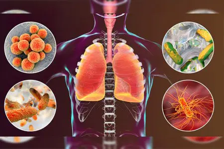
Most often, the following pathogens are detected in the biomaterial of patients with atypical pneumonia:
Legionella.
Mycoplasma.
Chlamydia.
Coronavirus.
A feature of all these microorganisms, in contrast to pneumococci (causative agents of ordinary pneumonia), is their ability to penetrate directly into the cells of the lung tissue, and beyond. The further vital activity of an individual pathogen determines the nature and severity of the disease.
Clinical picture in adults
A significant proportion of patients suffer from atypical pneumonia in mild or moderate form.
The first signs of infection are similar to the manifestations of SARS:
Bad feeling.
Feeling of weakness, weakness.
Headaches, muscle pains.
Dryness of mucous membranes.
Cough.
Pharyngitis.
Rhinitis.
Laryngitis.
When listening to the lungs, weakened breathing, crepitus, single wheezing are recorded. In laboratory blood tests – an increase in ESR, leukocytosis.
A hallmark of SARS is a characteristic paroxysmal cough. Usually cough occurs when the patient assumes a certain position.
Symptoms depending on the pathogen
The clinical features of SARS, the risk of complications and the prospect of recovery depend on the type of pathogen. This fact is important for determining the therapeutic tactics and determines the success of the treatment.
Mycoplasma pneumonia
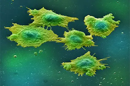
Infection with mycoplasma (Mycoplasma pneumoniae) is carried out in close interaction with a sick person. When clarifying the epidemiological anamnesis, it most often turns out that the patient was in some closed room among a large number of people.
Features of the flow. Atypical pneumonia of mycoplasmal origin is most often mild. A severe course is observed in patients with other serious pathologies, reduced immunity.
Signs. The first clinical manifestations are similar to influenza symptoms appear 3-11 days after infection. Patients complain of weakness, unproductive hacking cough, dry mucous membranes. Later, thick sputum joins, sometimes with an admixture of purulent discharge. The active effect of mycoplasma causes muscle pain in the back, hips, joint pain in the form of polyarthritis. Nosebleeds, rashes in the form of blisters, spots, papules are often observed.
Weather. Atypical mycoplasmal pneumonia has a favorable prognosis. Among the possible complications, the greatest danger is bronchiectasis, bronchiolitis, pneumosclerosis.
Chlamydial pneumonia
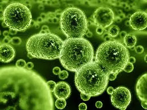
Chlamydia (Chlamydophila pneumoniae) infect the body by aerosol. The incubation time can be delayed up to 30 days.
Features of the flow. The disease is characterized by a sudden increase in temperature to 38-39 ° C. On the first day, symptoms of an acute viral infection appear. In 80% of cases, the infectious-inflammatory process affects both lungs.
Signs. Patients complain of muscle and joint pain, shortness of breath, cough with viscous sputum, hypertrophy of the cervical lymph nodes.
Weather. Chlamydial pneumonia is characterized by a long, but not severe course, however, due to the long stay of the bacterium inside the body, it leads to an allergy to chlamydia antigens. Possible complications such as bronchial asthma and obstructive bronchitis.
legionella pneumonia
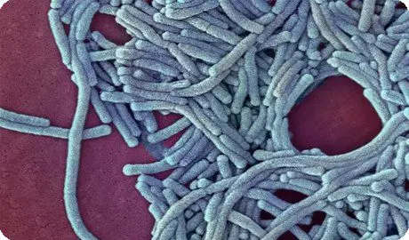
Legionella pneumonia pathogen (Legionella pneumophila) is found in air conditioning or water supply systems, in ultrasonic humidifiers, in humidifiers of ventilator systems. Infection is carried out by aerosol. The incidence is much higher during the summer months.
Features of the flow. From the fact of infection to the appearance of the first symptoms, it takes from 4 to 10 days. A specific sign of the disease is the persistence of a high temperature up to 40 ° C for several days.
Signs. Patients complain of chills, headaches, dry cough. Over time, mucopurulent sputum joins. Possibly hemoptysis. The disease is complicated by shortness of breath, pain in the pleura, dyspeptic symptoms, tachycardia, nausea, diarrhea.
Forecast – unfavorable. The death of the patient can occur as a result of complications, a combination of respiratory and renal failure.
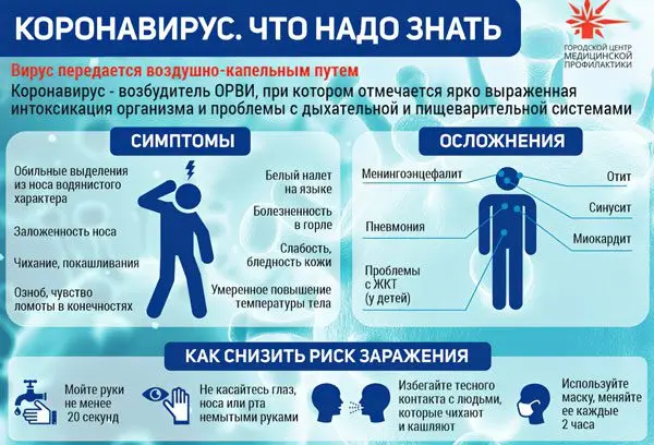
Coronaviruses of the Coronaviridae family have a negative effect on the lower respiratory tract. Infection occurs by aerosol from a sick person or an asymptomatic carrier.
Signs. The first clinical symptoms are recorded on the 2-10th day. Patients have a high temperature up to 38-40 ° C, chills, increased sweating, headaches. Over time, a dry cough without sputum production, shortness of breath joins. The patient’s condition is aggravated – respiratory dysfunction develops, a disorder of cardiovascular activity, signs of general intoxication.
Forecast – unfavorable, since the disease is characterized by high mortality. The patient dies as a result of toxic infectious shock, respiratory distress syndrome, acute respiratory and cardiovascular failure.
Features of the course of the disease in children
The child’s body does not always respond to inflammation in the lung tissue with an increase in body temperature.
Against the background of a normal or slightly elevated (subfebrile) temperature, the child’s well-being quickly worsens, specific symptoms of the disease appear:
Lethargy, drowsiness.
Lack of interest in food.
Difficulty breathing, shortness of breath.
Attacks of profuse sweating.
Vomiting.
Liquid, multiple stools.
In the case when the primary cause of the disease is mycoplasma, palpation is determined by an increase in the boundaries of the liver, spleen, polymorphic rashes are visible on the skin. The child takes a forced position – lies on the side of the affected lung. This position brings relief, reduces soreness in the chest. With the course of the disease, the child’s condition worsens – there is a change in the depth of breathing, a disorder of respiratory activity. In some cases, systematic respiratory arrests are observed – short-term apnea.
A sufficient immune response contributes to the recovery of the patient. With reduced protective properties of the body, the disease progresses. Against the background of the worsening symptoms of SARS, complications develop – severe acute respiratory syndrome (SARS), acute respiratory disease syndrome (SARS). The most common cause of death is acute respiratory failure.
Diagnostics
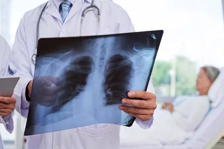
Diagnosis of SARS is characterized by an integrated approach. It is mandatory to complete all stages of the examination:
Assessing the severity of symptoms.
Examination of the patient. Auscultation revealed moist rales and crepitus.
Physical examination – X-ray, MRI or CT of the chest.
Laboratory tests of blood and urine.
Exciter type verification.
X-ray examination
When the first symptoms appear, indicating the development of SARS, chest X-ray provides the necessary data for an accurate diagnosis. CT or MRI methods are used much less frequently, as a rule, in cases where the interpretation of images is difficult, it is not possible to establish changes on the plain radiograph.
The description of the obtained images is carried out by the radiologist. In conclusion, the nature of the changes must be indicated – the type of infiltration, the definition of pleural effusion, darkening.


