Contents
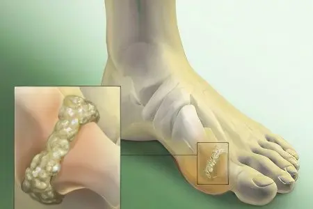
Medical science does not single out the deposition of salts in the joints as a separate disease. Rather, it is a simplified name for growing osteophytes, as a result of which the bone begins to deform, and pathological disorders in the functioning of the musculoskeletal system are formed.
Causes of salt deposits in the joints
The deposition of salts in the joints occurs as a result of a malfunction of metabolic processes, which is facilitated by:
irrational nutrition, which includes spicy meat dishes;
alcohol abuse;
smoking;
maintaining a sedentary lifestyle;
hypothermia of a particular joint or the whole body.
The disease develops quite slowly, but early diagnosis allows you to cope with the disease in a shorter time and with maximum efficiency.
Symptoms of salt deposits in the joints

Several symptoms can be identified:
Symptoms of the disease, first of all, are accompanied by the appearance of a specific “crunching” sound in the joints.
In some cases, movement may cause pain in the affected joint.
Gradually, the movement of the joint becomes more limited up to complete immobilization.
The joint is deformed.
Salt deposits in the knee and shoulder joint
A disease in which there is a deposition of salts in the knee joint is called gonarthrosis. It is this joint that, according to statistics, is most often affected.
The disease is accompanied by severe pain not only when walking, but also at rest. Untimely seeking medical help leads to irreversible consequences.
The main signs of gonarthrosis include not only painful sensations that increase with a load on the joint, but also “tightness” of the skin and stiffness of the joint in the affected area; long “pacing” after sleep or prolonged sitting; crunches when bending the lower limb; swelling and swelling of the knee; in a more severe degree of the disease, it is impossible to fully straighten or bend the leg.
The pathogenesis of the disease is represented by two main forms: primary (or idiopathic), characteristic of the elderly, and secondary, arising from an already existing pathology of the knee joint, for example, as a result of a mechanical injury. The primary type of gonarthrosis is most often bilateral, while in the secondary, in most cases, a unilateral process is observed.
It is customary to distinguish 3 stages of gonarthrosis:
The first stage, which is characterized by the initial manifestations of the disease (periodically occurring dull pain in the knee, especially after a load on the joint, there is some swelling of the affected area). There is no deformation at this stage.
The second stage It is manifested by an increase in the manifestations of the symptom complex of the previous stage: pain sensations appear more often and become more intense and prolonged. When moving, crunches and clicks appear in the joints. There are some restrictions in flexion and extension of the lower limb. The joint increases in volume, primary signs of deformation appear.
The third stage – this is the highest degree of manifestation of all the symptoms of the previous stages. Pain is present almost constantly – both during movement and at rest. Walk is disturbed. Restrictions on motor activity become pronounced. Joint deformity is visualized and proceeds in X- or O-shape.
The prognosis of the disease is not favorable. Most often, the affected joint is completely deformed, and the disease leads to disability.
Periarthritis should be understood as a disease accompanied by the deposition of salts in the shoulder joint. In most cases, one shoulder is affected or the degree of damage on one side is significantly greater than on the other.
The initial stage of the disease is characterized by the appearance of pain during physical activity, especially when it becomes necessary to raise your hands up. In the beginning, such pains appear irregularly and do not attract much attention. But gradually the discomfort becomes constant, causing inconvenience even during sleep.
If an inflammatory process develops in the affected joint, swelling may appear. Joint mobility is limited. There are characteristic “crunchy” sounds and clicks.
The prognosis of the disease is quite favorable. There may be cases when treatment is not required at all, but this is determined by the doctor. Complications of periarthritis occur in extremely rare cases.
calcium gout

Inflammation of the joints can occur as a result of the deposition of calcium pyrophosphates. In this case, they speak of such a phenomenon as calcium or false gout. A similar disease is accompanied by arthritis in the ankles, hips or hands.
The main symptoms of the disease include spontaneously arising and in the same way disappearing pain (quite strong) and swelling.
The causes of false gout lie in the following provoking factors: advanced age, the presence of hypothyroidism, hemophilia, manifestations of amyloidosis, heredity.
The correct delivery of the diagnosis requires a laboratory study of the joint fluid. The disease is confirmed by the detection of calcium porophosphate in the composition of the taken material.
Diagnostics
Diagnosis of salt deposits in the joints includes several special methods:
radiography is a reliable method, but with its help the disease can be diagnosed only after 5 years of the course;
arthroscopy – includes the procedure of using an arthroscope, an apparatus that is inserted into the affected joint through a micro-incision. Thus, the doctor has the opportunity to see the state of the joint from the inside;
computed tomography – a method by which the size of the joints, their qualitative characteristics and pathological processes associated with the growth of cartilage and the appearance of osteophytes are determined;
magnetic resonance diagnostics – allows you to determine the layered structure of the joint and bones, soft tissues and pathological formations;
thermography – an auxiliary research method that shows thermographic data, including index, temperature gradient and asymmetry of the joints;
Diagnosis and subsequent treatment is impossible without a thorough laboratory study, including uric acid analysis, erythrocyte sedimentation rate, leukocyte count, Zimnitsky test, study of fluid puncture (in the case of knee joint disease), etc.
Treatment
Medical science in the treatment of salt deposits in the joints relies on an integrated approach:
Non-steroidal anti-inflammatory drugs can reduce and even completely relieve pain and inflammation in the joints.
With the help of hormonal drugs – corticosteroids – acute attacks are controlled. Uricosuric dosage forms with the deposition of salts in the joints help maintain the level of uric acid at the normal level. Such drugs are prescribed only if all the necessary tests have been carried out and their qualified assessment by a specialist has been given.
Treatment of shoulder periarthritis It will be more successful if the appeal for medical help was received at the first signs of the disease. A neglected form will require more time and effort to defeat the disease.

When the development of periarthritis began with the displacement of the intervertebral joints, then manual therapy becomes an effective method of treatment. Its main task is to eliminate bias.
If the disease is associated with impaired blood circulation in the shoulder region as a result of myocardial infarction or surgical manipulations on the mammary gland, then special angioprotective drugs are necessarily present in the treatment. Their main task is to improve blood circulation.
In the case when the deposition of salts is accompanied by liver diseases, it becomes necessary to follow a diet and take special enzymatic agents to restore its functions.
With direct treatment of the tendons of the shoulder non-steroidal anti-inflammatory drugs, compresses are prescribed. In some situations, the use of laser therapy is useful.
Hirudotherapy. The effectiveness of the treatment of shoulder periarthritis through medical leeches – hirudotherapy has been proven. Significant improvements appear already at 5-6 sessions. But this method must be used with extreme caution, because. with this disease, an allergy to the use of leeches often develops.
Corticosteroids. Good results are obtained by conducting a cycle consisting of 2-3 periarticular injections of corticosteroid hormonal preparations. A mixture of hormones and an anesthetic is injected into the affected tendon or into the region of the periarticular synovial sac. Such a procedure is not capable of leading to a complete recovery, but it alleviates the condition of patients in more than 80% of cases. To enhance the effect of injections, they must be combined with a set of therapeutic measures such as post-isometric relaxation and special exercises that improve the mobility of the capsular part of the joint.
Postisometric relaxation represents the most successful way to treat salt deposits in the shoulder joint. The course consists of 12-15 sessions. According to statistics, about 90% of patients, even with the most advanced form of the disease, recover as a result of using this method. The effect is enhanced by the parallel use of a laser, therapeutic massage, sessions of soft manual therapy (the so-called joint mobilization method), as well as if the course of post-isometric relaxation begins 2-3 days after the periarticular injection of corticosteroid hormones.
Early stage of gonarthrosis treated with conservative methods. Therapeutic procedures include physiotherapy, physiotherapy and massage sessions, the possibility of spa treatment, exercise therapy and the use of symptomatic medications. At the same time, taking medications is not intended to restore the function and structure of the joint or cartilage, but only to eliminate pain.
After the positive dynamics and improvement are noted as a result of the use of medications, manual therapy and a course of therapeutic massage are prescribed. With the help of such events, the muscles of the knee are strengthened, blood circulation and nutrition of the cartilage are corrected, and, accordingly, the position of the bone.
Launched stages diseases require surgical intervention. Knee arthroplasty is a surgery for the treatment of gonarthrosis. The procedure lasts an average of 90 minutes and is accompanied by minor blood loss. Materials for the manufacture of implants that replace the knee joint are highly durable and perfectly take root in the human body. The most common are stainless steel or titanium alloy, as well as ceramics and polyethylene – heavy-duty plastic.
The modern design of the implant is represented by the femoral, tibial elements and the patella prosthesis. Depreciation between them creates a special gasket made of polyethylene.
Treatment of salt deposits in the joints is a complex and lengthy process that requires regular use of medications. Along with this, it is necessary to reconsider the lifestyle, first of all, to follow a diet and give up bad habits.
To date, there are no clearly defined preventive measures to prevent the deposition of salts in the joints. This disease affects everyone without exception. However, a reasonable lifestyle that harmoniously combines proper nutrition and physical activity can become the key to the health of the human skeletal system.


