Contents

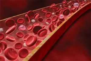
Reticulopenia is a decrease in the level of reticulocytes in the blood. Normally, their number is from 0,2 to 1,2% of the total number of red blood cells. Reticulocytes are young forms of red blood cells that contain granular inclusions.
Reticulopenia is indicated not only by a decrease in the number of reticulocytes below 0,2%, but also by their complete absence in the blood. Reticulopenia can develop in both an adult and a child. The absence of immature reticulocytes in human blood is not a variant of the norm and often indicates serious diseases, for example, anemia or bone marrow damage by metastases of cancerous tumors.
Causes of reticulopenia

The reasons for the decrease in the level of reticulocytes in the blood can be as follows:
Aplastic or hypoplastic anemia. Aplastic anemia is characterized by inhibition of the function of the red bone marrow, which naturally leads to a decrease in the number of reticulocytes in the blood.
Iron-deficiency anemia. Reticulopenia develops in moderate to severe disease.
B12 deficiency anemia. Lack of vitamin B12 in the body negatively affects the function of the bone marrow, so the maturation of red blood cells is disturbed.
Thalassemia leads to reticulopenia, since this disease damages the structure of red blood cells, with the destruction of their cells and the development of hemolytic crises. Thalassemia is a hereditary pathology.
Sideroblastic anemia.
Damage to the bone marrow by metastases of a cancerous tumor of a different localization.
Radiation sickness or radiation therapy. At the same time, young forms of all blood cells, and not just erythrocytes, will be absent in the bone marrow.
Autoimmune diseases, involving the organs of the hematopoietic system in the process.
Diseases of the kidneys. The kidneys have a direct influence on the processes of hematopoiesis. If there is any serious damage to these organs, then the production of erythropoietin is disrupted. Without erythropoietin, normal erythropoiesis is impossible, which leads to a decrease in the number of reticulocytes in the blood. Of particular danger to the hematopoietic system is renal failure, as an acute course. In this case, inhibition of erythropoiesis occurs, and the life span of erythrocytes is significantly reduced. This also applies to their young forms – reticulocytes.
Alcoholism. With alcoholism, the body as a whole suffers, and the bone marrow in particular. Naturally, the renewal of blood cells in such conditions is simply impossible.
Anemia Addison-Birmer (relapse of the disease). This is pernicious anemia, accompanied by a lack of vitamin B12 and folic acid. With this disease in humans, the number of reticulocytes first increases in the blood, and the relapse of the pathology is characterized by a decrease in all immature blood cells.
Myxedema. This disease develops against the background of a deficiency in the body of thyroid hormones. The blood picture proceeds according to the type of hypochromic anemia.
Symptoms of reticulopenia

By itself, reticulopenia does not have any symptoms. This condition develops only under the influence of a number of pathological causes. Therefore, it is precisely the symptoms of the underlying disease that come to the fore.
The main symptoms of aplastic anemia, which causes reticulopenia, are dizziness, severe weakness, fainting, shortness of breath, chest pain. There is an increased tendency to bleed. The human immune system becomes weakened, which increases the likelihood of developing infectious and purulent processes.
Iron deficiency anemia will be indicated by increased fatigue, drowsiness, dizziness, dryness and pallor of the skin, brittle nails and hair, inflammation in the oral cavity. A person becomes emotionally unstable, his memory and attention deteriorate, he begins to get sick more often.
Symptoms such as weakness, malaise, headaches, dyspepsia, and tinnitus indicate B12-deficiency anemia. A person has a tendency to become obese. The disease most often develops smoothly and is not noticeable to a person.
Symptoms of thalassemia depend on the type of disease. In general, they come down to bone pathologies, enlargement of the spleen and liver in size, pallor of the skin and other anemic manifestations.
With sideroblastic anemia, all the characteristic anemic symptoms develop: pale skin, increased fatigue. This disease is associated with a risk of developing diabetes mellitus, arrhythmias, and pulmonary insufficiency. Possible transmission of the disease by inheritance. In this case, her symptoms will disturb the child from childhood.
With the penetration of metastases into the bone marrow, in addition to reticulopenia, the patient will experience anemia, frequent dizziness, weakness increases. The pain extends to the lower back and ribs, to the pelvic bones. As the pathological cells grow, the pain sensations will intensify. A person will begin to lose weight, often get sick.
Radiation sickness affects all organ systems. A person develops a headache, weakness increases, body temperature rises, diarrhea can manifest. Often, blood pressure drops sharply, which leads to loss of consciousness.
In acute renal failure, which leads to reticulopenia, the amount of urine excreted sharply decreases. The patient develops diarrhea, vomiting and nausea, he becomes drowsy, may fall into a coma.
With myxedema, a person has dryness and pallor of the skin, increased swelling of the face and extremities. Hair becomes dry and begins to fall out. Often there is a low body temperature, blood pressure drops, cholesterol levels rise.
Diagnosis of reticulopenia

Three diagnostic methods can be used to calculate the level of reticulocytes in the blood:
Determination of the content of reticulocytes in the blood in a smear after adding a special dye to it. This is the simplest and cheapest diagnostic method. It is available to any laboratory. For staining reticulocytes, alkaline reagents can be used, for example, azure solution, which makes it possible to reveal the granular structure of young erythrocytes. Then the doctor counts their number under a microscope.
Determination of the content of reticulocytes in the blood using fluorescent microscopy. This is an accurate technique that requires special dyes and a fluorescent microscope in the laboratory.
Determination of the level of reticulocytes using modern hemolytic analyzers. This method allows not only to identify the relative percentage of reticulocytes in the blood, but also to provide additional information to the doctor regarding the quantitative content of cells of various forms (immature fractions, cells with high, medium and low RNA content).
Any of the above methods will require a blood donation. Some factors can distort the results of the analysis:
Wrong choice of anticoagulant.
Prolonged pressure on the limb with a tourniquet.
The patient is taking sulfonamides.
A recent red blood cell transfusion procedure.
Hemolysis of a blood sample.
All these factors must be taken into account when conducting research. Additional diagnostic tests are needed to clarify the cause of reticulopenia.
Treatment of reticulopenia

There is no cure for reticulopenia. This condition will be stopped on its own after it is possible to get rid of the cause that led to a decrease in the number of reticulocytes in the blood.
Therefore, the doctor may prescribe the following therapeutic measures:
If the patient has aplastic anemia, then he is prescribed blood transfusions, immunosuppressive therapy. You can get rid of the disease thanks to bone marrow transplantation.
Treatment of iron deficiency anemia involves following a diet with the inclusion of animal products (red meat, eggs, liver) in the menu. You need to take supplements containing iron. Already after 3-4 days from the start of therapy, reticulopenia in a patient should be replaced by reticulocytosis. This will indicate that the treatment was chosen correctly. Over time, the level of reticulocytes in the blood will return to normal.
Treatment of B12 deficiency anemia involves parenteral administration of vitamin B12. In severe cases of pathology, red blood cell transfusion is possible.
Treatment for thalassemia varies, depending on the form of the disease. With small b-thalassemia therapy is not required. Although some children need blood transfusions from birth, iron and glucocorticoids if they develop hemolytic crises. Any form of the disease requires the intake of folic acid and B vitamins. If a donor is found, then patients with thalassemia require a bone marrow transplant.
With sideroblastic anemia, the patient is prescribed vitamin B6 for up to 2 months. Severe pathology requires a blood transfusion.
The presence of bone marrow metastases requires emergency treatment. The patient will necessarily need to undergo a course of chemotherapy, after which he is prescribed radiation exposure to the brain. Only an organ transplant can save the patient.
Treatment of radiation sickness requires placing the patient in a sterile box. He is given drugs that neutralize the effects of radiation. The most powerful detoxification therapy is carried out on the first day of radiation sickness. In addition to forced diuresis, transfusion of erythrocyte and platelet mass, plasmapheresis may be required. The body will recover for a long time, sometimes throughout life.
Treatment of acute renal failure should begin with the elimination of the cause that led to the disruption of the kidneys. It is necessary to replenish the volume of circulating blood, to normalize blood pressure. To stimulate diuresis, the patient is prescribed osmotic diuretics and Furosemide. Antibacterial drugs may be used as needed. Hemodialysis is an emergency treatment measure.
Treatment for Addison-Birmer anemia requires lifelong vitamin B12 and folic acid supplementation. Moreover, atrophy of the gastric mucosa leads to the fact that in tablet form these drugs may not be absorbed. Therefore, the patient is prescribed their parenteral administration.
Treatment of myxedema requires the use of thyroid hormones. In parallel, carry out therapy aimed at eliminating the symptoms of the disease.
Thus, a wide variety of treatment regimens may be required to normalize the level of reticulocytes in the blood. Therapy depends on the underlying disease and should be carried out only after a qualitative examination of the patient.


