Contents
Raynaud’s Disease – This is a disease from the group of angiotrophoneurosis, which is characterized by an acute violation of arterial circulation in limited areas of the body (in the feet and hands). The disorder develops as a result of the influence of exogenous and endogenous factors. Raynaud’s disease is also called angiotrophoalgic syndrome, vascular-trophic neuropathy, vasomotor neurosis.
The disease is more common in countries with cold climates. It affects women aged 5 to 40 50 times more often.
Raynaud’s disease should be distinguished from Raynaud’s syndrome, since despite the similarity of symptoms, they differ in etiological factor. The fact is that after Maurice Raynaud described the signs and etiology of the disease, it was found that it can develop as an independent disease due to impaired functioning of the central nervous system, and can act as a syndrome of some other pathologies. This is the reason for the difference between the two concepts.
Raynaud’s disease symptoms

Symptoms of Raynaud’s disease can be considered depending on the stage of the pathological process:
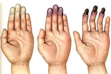
Symptoms of the first stage of Raynaud’s disease. The affected area – this may be the surface of the feet or hands, and sometimes the auricles, lips, nose, begin to turn pale, become cold when palpated. Sensitivity in the affected area disappears or is significantly reduced.
The duration of an attack varies widely and can take anywhere from a minute to several hours. As the spasm is removed from the vessels, the skin returns to its normal state.
The frequency of occurrence of attacks varies, but they will be repeated the more often, the more the disease progresses. Over time, in addition to a violation of sensitivity, a person will begin to experience pain.
Paresthesia, coldness of the fingers, whitening of the skin at this stage most often occur as a result of exposure to cold, due to excitement, smoking, etc.
Symptoms of the second stage of Raynaud’s disease (angioparalytic). At this stage, in addition to vasospasm, the phenomena of asphyxia join. During an attack, the skin acquires a blue-violet color, in parallel there are pronounced pains, sensitivity completely disappears. After the attack is completed, the skin on the affected areas becomes bright red, and a vascular pattern in the form of a grid may appear.
To start an attack at the second stage of the development of the disease, there is no longer any need for the influence of any factor, it occurs spontaneously and lasts for a long time.
Symptoms of the third stage of Raynaud’s disease (trophoparalytic). Asphyxia increases in time, as a result of which blisters appear on the affected bluish and edematous parts of the body. Inside them you can see bloody contents. When the bubble opens, areas of necrotic tissue are found under it. If the disease has a severe course, then the muscles can be damaged, up to the bone. Over time, the ulcerated surface is scarred.
The first and second stages can last from 3 to 5 years. If the affected area is the palms or the surface of the feet, then there is often a combination of symptoms of all three stages at the same time.
Raynaud’s disease has a tendency to relapse. The difference between the disease and the syndrome in terms of the severity of the symptoms lies in the fact that with the syndrome, gangrene of the affected area is more often observed, trophic disorders are more pronounced, areas of necrosis are formed, damage to the nail plates is attached, their deformation.
Causes of Raynaud’s disease
The causes of Raynaud’s disease cannot be considered separately from the mechanism of the development of the disease. It is based on violations of the organic and functional plan, affecting both the vascular walls and the apparatus responsible for their innervation. As a result, there is a violation of the nervous regulation of blood vessels, so they react to various effects with spasms, followed by increasing atrophy.
Causes of Rein’s Syndrome:
Autoimmune diseases affecting connective tissue: systemic lupus erythematosus, systemic scleroderma, rheumatoid nodosa, rheumatism, Sjögren’s syndrome, dermatomyositis, periarteritis.
Vascular diseases are Takasyau disease, obliterating atherosclerosis of the legs, etc.
Damage to the peripheral nerves on the background of diabetes mellitus (polyneuropathy).
Intoxication of the body with lead, arsenic salts, cytostatics and ergotamine.
Blood viscosity disorders: cryoglobulinemia, polycythemia vera, Waldenström’s macroglobulinemia.
Osteochondrosis of the upper thoracic and cervical.
Prolonged exposure to vibration during the development of vibration disease.
Lack of autonomic nervous regulation – syringomyelia.
Violations in the functioning of the adrenal glands, thyroid and parathyroid glands.
Rarely, Rein’s syndrome provokes accessory cervical rib syndrome, carpal tunnel syndrome, and scalenus anterior syndrome.
In turn, the causes of Raynaud’s disease lie in the pathologies of the central nervous system and spinal cord with the involvement of the hypothalamus, brain stem and cortex in this process. These pathological processes lead to the fact that the impulses that regulate the work of blood vessels are transmitted with violations.
Diagnosis of Raynaud’s disease

Diagnosis of Raynaud’s disease is reduced to the analysis of the patient’s complaints, and is also based on the collection of anamnesis. The primary role is played by an inadequate reaction with a change in the color of the skin of the feet and palms to the effects of cold. Asbestos whitening of limited areas of the skin under the influence of low temperatures is observed in 78% of cases in patients with Raynaud’s disease.
The following diagnostic criteria for evaluating the patient’s response to a cold test in the period between attacks have been identified:
The skin color does not change – the syndrome is not confirmed;
The skin changes color, there is a feeling of numbness, paresthesia – there is a possibility of a syndrome;
The skin changes color to cyanotic, followed by redness, the attacks are repeated – the syndrome is confirmed.
In addition, the following instrumental examination methods are used, such as: thermography of the affected area, Doppler flowmetry, angiography of the peripheral vascular bed, capillaroscopy of the vessels of the anterior surface of the organ of vision and the nail bed.
In order to identify Raynaud’s disease in a patient or Raynaud’s syndrome, the following differential signs are used:
Diagnostic evaluation criterion | Raynaud’s syndrome | Raynaud’s Disease |
Age of disease manifestation | From 30 and older | Doesn’t matter |
Presence of a systemic connective tissue disease | Lupus erythematosus, scleroderma, etc. | No |
Limb lesions are symmetrical | Yes | No |
Trophic changes, necrosis, tissue atrophy | There are | None |
Capillaroscopy results | There are morphological changes in the microvascular pattern | No changes |
ESR | Increased | Within normal limits |
ELISA of blood for antinuclear antibodies | Positive | Negative |
The presence of vascular spasms (crises) in the lung tissue and kidneys | There are | None |
Plethysmography on the background of a cold test | Pressure reduced | The pressure is normal, or slightly reduced |
Doppler flowmetry | The blood flow is greatly reduced | Within normal limits |
Raynaud’s syndrome is confirmed by the presence of other diseases for which it acts as a symptom complex. Raynaud’s disease is presented as a diagnosis if such diseases are absent, and also on the basis of a thorough diagnosis.
Raynaud’s disease treatment
The treatment of Raynaud’s disease primarily boils down to eliminating all possible factors that contribute to the occurrence of spasms. These are smoking, hypothermia, exposure to vibration, as well as other exogenous influences.
It is important to establish the underlying disease that provoked Raynaud’s syndrome, if such a diagnosis is confirmed. Depending on this, a person will be assigned a certain disability group. However, if the patient is unable to work, then disability may be assigned due to Raynaud’s syndrome.
Emergency assistance during an attack of vasospasm is reduced to the following activities:
To protect a person from the influence of a provocateur factor as much as possible.
Warm the affected area – perform a massage, offer the patient a plentiful hot drink.
Make an injection or take an antispasmodic drug orally (Platafillin, No-shpu, Drotaverin). For severe pain, an analgesic should be taken.
At the discretion of the attending physician, the following drugs are prescribed:

Nifedipine-based vasodilators (Nifedipin, Cordaflex, Osmo-adalat, Corinfar, Cordipin, Nifecard, Fenigidin), as well as Nicardipine and verapamil-based agents (Isoptin, Finoptin, Verogalid).
Drugs – angiotensin-converting enzyme inhibitors (Capoten, Captopril).
Ketanserin, as a blocker of the effects of serotonin.
Preparations for the normalization of blood composition, to improve its microcirculation – Trental, Agapurin, Pentoxifylline, Dipyramidol, Vasonite.
Preparations from the group of lipid physiologically active substances – Vap, Vazaprostan, Alprostan, Caverject.
It is mandatory to supplement conservative therapy with physiotherapeutic methods of treatment. Well proven procedures such as: galvanic baths, mud therapy, UHF, hyperbaric oxygen generation, reflexology. On the recommendation of a specialist, a complex of exercise therapy should be performed.
If conservative methods do not give an effect, then it is possible to perform an operation – sympathectomy, or ganglionectomy.
Progressive methods of treating Raynaud’s syndrome include stem cell therapy, which contributes to the normalization of blood flow.
The prognosis for life is generally favorable, however, it largely depends on the progression of the underlying disease. In addition, spontaneous cessation of ischemic attacks is possible with a change in lifestyle, after quitting smoking, with a change in climate and working conditions.
There are no primary measures to prevent Raynaud’s disease, and secondary preventive measures are reduced to the maximum elimination of provoking factors.


