


By the term “precancerous condition” is meant a skin lesion that likely precedes the development of skin cancer. Among the many different units, the ones described below are the most common.
Types of precancerous conditions and skin cancers
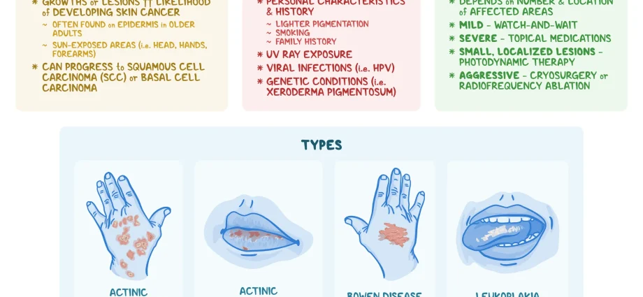
1. Senile, or solar keratosis
It is most often located on the face, baldness, edges of the auricles, backs of the hands. Initially, the lesions of actinic keratosis are hardly visible, they are more often felt by touch as rough spots. Later they are marked as brown foci, covered with strongly adherent rough scales. Post-exfoliation lesions should undergo electrocoagulation or cryotherapy.
2. White keratosis
These are changes on the mucous membranes of the mouth, as well as the genitals. The latter are associated with infection with papillomaviruses with carcinogenic features. In the oral cavity, the appearance of white keratosis is favored by cigarette smoking, as well as by long-term trauma to the mucosa associated with tooth damage. White keratosis is initially porcelain-white spots, then papules, possibly with erosions, and finally white papillary growths. In addition to removing the above-mentioned causative factors, cryotherapy is used in keratosis.
3. Solar cheilitis
The lips, especially the lower lip, are most vulnerable to solar cheilitis. Multiple exposure (farmers, anglers, etc.) causes:
- edema,
- dryness,
- exfoliation,
- sometimes painful cracks,
- finally, foci of infiltration and papillary hyperplasia.
Prevention
The prophylaxis of solar red lips consists in the use of special light-absorbing lipsticks or lip creams.
Treatment
The resulting changes are treated with anti-inflammatory and exfoliating creams, if necessary with electrocoagulation.
4. Seborrheic wart
It is a mild epithelial hyperplasia. It occurs on the torso, face, neck, back of the hand. Changes occurring in the course of a seborrhoeic wart may be single or multiple, they are sharply demarcated from healthy skin, light or dark brown, raised (papular), with a rough, uneven surface. Sometimes the horny masses on the surface of the efflorescence may crumble by themselves, which causes the lesion to temporarily flatten, but after some time it grows back again. The lesion is not malignant and does not become malignant.
Treatment
In the treatment of seborrheic warts, cryotherapy, electrocoagulation or lesion excision are used.
5. Skin cancers
The skin has basal cell carcinoma and squamous cell carcinoma. The former is much more common (twenty cases per 100 population), while the latter is 10 times less common.
6. Basal cell carcinoma
It has a relatively milder course than squamous cell carcinoma, does not metastasize in principle, but is highly destructive to tissues. The most common locations are the nose (especially the side of the nose), the angle between the nose and the lower eyelid, the pre- or behind-the-ear area, which does not mean the cancer cannot appear anywhere on the skin.
symptoms
The first lesion occurring in basal cell carcinoma is a small nodule approx. 3-4 mm in diameter, flesh-colored, painless. With time, the lesion enlarges and creeps. It leaves an ulcer or scar inside, a lump with an uneven surface or a dark color.
In people with neglected ailments, tissue breakdown and damage can be very great.
Diagnosis
Diagnostics is based on taking a skin specimen and histopathological examination.
Treatment – the most effective and in all respects the best method is surgical removal of the lesion (simple excision of a small lesion or plastic resection in the case of large cancer).
7. Squamous cell carcinoma
It is more malignant than basal cell carcinoma and metastasizes (mainly to the lymph nodes). The most common location of lesions is on the border of the skin and mucous membranes, so e.g. the lower lip, genitals, but it can appear anywhere on the skin, especially on the basis of precancerous conditions. This cancer appears as a nodule or lump with a papillary surface or as a hard-based ulcer. It is painless. It may be accompanied by enlargement of the surrounding lymph nodes.
Diagnosis – is made on the basis of the cut from the lesion. Treatment, in turn, is primarily a surgical procedure.
The content of the medTvoiLokony website is intended to improve, not replace, the contact between the Website User and their doctor. The website is intended for informational and educational purposes only. Before following the specialist knowledge, in particular medical advice, contained on our Website, you must consult a doctor. The Administrator does not bear any consequences resulting from the use of information contained on the Website.