Contents
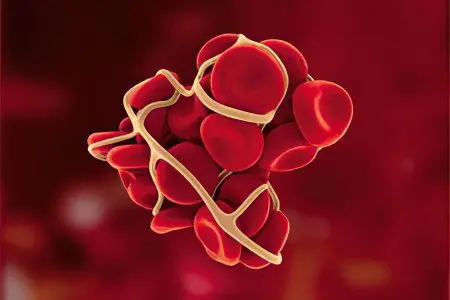
Platelet aggregation is the concentration of certain blood cells (Bizzocero’s plaques) to eliminate damage to the vessel, accompanied by blood loss. Violation of integrity with minor damage to small vessels against the background of normal hemostasis does not threaten massive blood loss. Light bleeding, according to many people, spontaneously stops after a short time. Not everyone knows that in this complex process of preventing significant blood loss, a lot depends on platelet aggregation.
Platelet aggregation or natural stop of bleeding
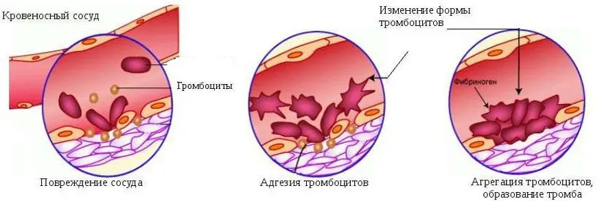
The process of stopping bleeding in the vascular network (capillaries, venules, arterioles) goes through several stages:
After damage to the vessel, its spasm occurs, which makes it possible to partially reduce the intensity of bleeding.
At the site of injury to the vascular wall, blood plates are concentrated, which partially cover the defect of the damaged area – platelet adhesion occurs.
At the site of the vessel defect, platelets accumulate, forming conglomerates, this is platelet aggregation, the first stage of thrombus formation.
As a result of irreversible aggregation, a platelet plug is formed. It is loose, does not hold firmly on the wound, with a slight mechanical impact on it, bleeding resumes.
Under the influence of thromboplastin of fibrin threads, the blood plug acquires density, shrinks, retraction of the thrombin thrombus occurs, and blood loss stops.
The picture below shows the stages of thrombus formation:
Platelet aggregation when bleeding stops is not the final stage of an important process, but this does not make it less important. This phenomenon, which is extremely important in stopping bleeding, has a downside. With increased platelet aggregation, platelets, even in the absence of bleeding, stick together, forming blood clots. These clots, moving through the blood vessels, provoke their blockage, disrupting the blood supply to the organs.
This is how myocardial infarction, pulmonary infarction, kidney infarction, ischemic stroke of the brain occur. In these cases, active antiplatelet therapy is prescribed for the prevention and treatment of thrombosis.
A seemingly insignificant pathological reaction of spontaneous platelet aggregation can lead to thromboembolism of the leading arteries, and even to the death of the patient.
What does platelet aggregation say in a blood test?
To study the ability of platelets to aggregate in the laboratory, an imitation of natural conditions is created – the circulation of blood cells in the bloodstream.
This study is carried out on glass with the addition of inducing substances that normally participate in similar reactions in the human body:
adrenaline;
Collagen;
ADP;
Thrombin.
As an inductor, ristomycin (ristocein) is additionally used, which has no analogues in the human body. Each inducer of platelet aggregation has its own normal range, which is slightly different in different laboratory conditions.
The rate of platelet aggregation depending on the inductor of the reaction
Method for determining platelet aggregation | Norm,% |
ADP blood test | 30,8 – 77,8 |
Blood test with adrenaline | 35,0 – 92,5 |
Collagen blood test | 46,5 – 93,2 |
Blood test with ristomycin | 58 – 167 |
For the diagnosis of cardiovascular pathologies, the determination of spontaneous platelet aggregation is of great importance, when the plates of blood cells stuck together circulate through the circulatory system.
Violations in the microcirculation zone:
Changes in the vascular walls in the microcirculation zone;
The formation of aggregates from platelets, which increase the risk of developing pathology of the heart and blood vessels, serious complications.
Methods for determining the parameters of the CAT:
Change in the optical density of the platelet suspension;
Visual assessment of platelet aggregation.
In the laboratory, for the diagnosis of pathological platelet aggregation, the following are used:
Optical aggregometers – register platelet aggregation in plasma;
Conductometric aggregometers – measure the level of aggregation in whole blood.
The data obtained is displayed as a curve on the aggregation chart. The method of laboratory diagnostics of the level of platelet aggregation is reliable, but requires high labor intensity, a large volume of blood plasma.
Norm and deviation during pregnancy
Deviations from the norm in the level of platelet aggregation in any direction is a sign of pathology. During pregnancy, a blood test is required to determine the risk of thrombosis. In the last trimester of pregnancy, the female body prepares for possible blood loss by moderately increasing blood clotting. If platelet hyperaggregation is detected, this condition must be corrected.
You can clarify the normal range in the laboratory, where the specialist will report on the risk of increased or decreased intensity of aggregation, on the likelihood of negative complications of pregnancy.
Platelet aggregation with inducers
According to the standard, for a more accurate diagnosis of the process, a blood test to determine the level of platelet aggregation is carried out with at least 4 inducers.
Inductor ADF
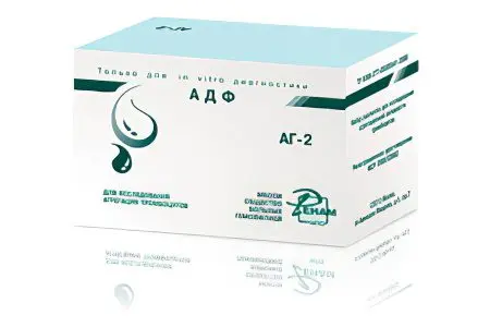
Carrying out diagnostics with ADP allows you to identify a process failure in the following diseases and conditions:
Ischemia, myocardial infarction;
Atherosclerosis;
Diabetes;
Arterial hypertension;
Violation of cerebral circulation;
Hyperlipoproteinemia;
Thrombopathy of hereditary origin;
Thrombocytopathy in hemoblastosis;
Taking drugs that inhibit the activity of platelets.
Diseases that provoke a decrease in the level of platelet aggregation:
Essential athrombia – a violation of the functionality of platelets;
Wiskott-Aldrich syndrome is a rare genetically determined disease that occurs depending on the sex of the patient, is associated with changes in the size and shape of cells;
Glanzman’s thrombasthenia is a genetic disease expressed in the absence of receptors for fibrinogen and glycoproteins;
Thrombocytopathy in uremias;
Aspirin-like syndrome – a violation of the second phase of platelet aggregation;
Secondary disorders of platelet aggregation in hemoblastosis, hypothyroidism, therapy with antiplatelet agents, NSAIDs, diuretics, antibacterial drugs and blood pressure lowering agents.
Diseases that provoke an increase in the level of platelet aggregation:
Activation of the coagulation system during psycho-emotional stress, the formation of immune complexes, taking certain medications;
resistance to aspirin;
Viscous platelet syndrome: increased level of aggregation, predisposition to adhesion.
The inducer is collagen
Going beyond the limits of normative indicators in the reaction with the use of collagen is diagnosed with violations at the stage of adhesion. The decrease in the level of platelet aggregation has the same reason as in the ADP tests. An increase in the level accompanies vasculitis, viscous platelet syndrome.
Inductor with adrenaline
The study of indicators of platelet aggregation in the adrenaline test is considered the most informative diagnostic method. It fully shows the internal mechanisms of activation, including the “release reaction”. The decrease in the normative indicator is typical for similar causes found in the reaction with ADP and with collagen. An increase in the intensity of platelet aggregation is associated with increased viscosity of blood platelets, with stress, with the intake of certain drugs.
Ristocetinoma inducer
The study is carried out when diagnosing von Willebrand syndrome. The study of ristocetin-cofactor activity of platelets helps to detect the severity of this factor.
All types of diagnostics using aggregation inductors make it possible to objectively assess the functionality of platelets. Another purpose of diagnostics is to evaluate the effectiveness of the use of antiplatelet agents, help in selecting the dosage of drugs.
Additional Information
The adhesion of platelets, the formation of a hemostatic plug, involves complex methods of platelet activation and the reactions that follow.
What is the role of platelets?
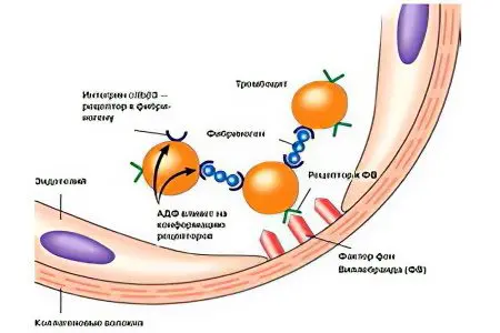
Functions of blood cells in the implementation of vascular-platelet hemostasis:
Providing angiotrophic function to maintain the normal structure and functionality of the vascular walls of small venules and arterioles.
Ensuring the adhesive-aggregation function, which consists in the concentration of platelets and their gluing to the damaged basement membrane of the vessel (adhesion), the formation of a hemostatic plug (aggregation), which allows you to stop bleeding in a short time.
Maintaining spasm of damaged capillaries to prevent increased blood loss.
Participation in coagulation processes, in fibrinolysis reactions.
The main stimulator of platelet plug attachment is collagen, although other components of the connective tissue can also perform these functions.
Changes in the appearance and functionality of platelets during bleeding
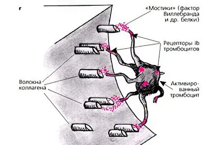
When a vessel is injured, platelet preparation for response begins long before arrival at the emergency site:
They change their shape from flat disc-shaped to spherical, acquire pseudopodia. These processes are designed to bind to each other and attach to the basement membrane of the vessel.
Arriving at the emergency site, the blood plates are ready to activate adhesion and aggregation, they attach to the walls of the vessel in less than 5 seconds.
Platelets located in the space of the circulatory system are concentrated into conglomerates of 3-20 cells.
Conglomerates that arrived at the site of damage are combined with platelets that are primarily adherent at the site of exposure of the basement membrane.
Cell concentration as a complex biochemical process
In the process of adhesion and aggregation of platelets, external and internal factors are involved:
Energy costs;
reaction stimulants;
Restructuring of cells.
For example, platelets cannot perform their functions without a glycoprotein, a plasma cofactor for collagen (Willebrand factor). It is produced in the vascular walls, and platelets, moving through the veins and arteries, deposit it for the future in their granules to be released if necessary.
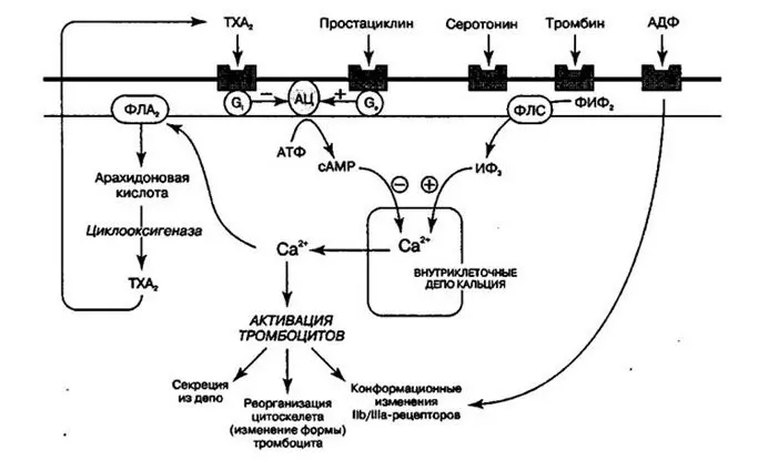
Platelet aggregation stimulators that turn on in activation mode:
Collagen is the most significant stimulant.
ADP – in the initial stage of aggregation is released from the damaged basement membrane of the vascular wall and from erythrocytes in the area of injury, then it is released by platelets that have passed through primary adhesion and activation.
Adrenaline and serotonin – activate platelet membrane enzymes, promote the formation of arachidonic acid and thromboxane, which intensively activate the reaction.
Prostaglandins – when activated, they form thromboxane in smooth muscles, at the last stage of aggregation they ensure the entry of prostacyclin into the blood, which suppresses the activity of platelets that is no longer necessary.
Thrombin – helps to strengthen and increase the strength of the hemostatic plug.
Focusing on the mechanism of platelet hemostasis reaction, one can understand the etiology of diseases associated with impaired blood clotting.
Vulnerability of blood cell concentration

Separate endogenous and exogenous factors provoke pathological processes in the mechanism of platelet hemostasis.
The most vulnerable place is the “release reaction”, when there is no connection and gluing of platelets, no hemostatic plug is formed.
For the qualitative implementation of the blood coagulation process, the presence of proteins is necessary: fibrinogen, albumin, gamma fraction, as well as phospholipid factor, Ca2+, Mg2+. Proteins create a “plasma atmosphere” for the full functioning of platelets, but some derivatives of protein cleavage can inhibit the aggregation of blood plates, especially those that were obtained from the breakdown of fibrinogen and fibrin.
If all participants in platelet hemostasis perform their functions, the aggregation of these blood cells is able to stop bleeding, but in large vessels where blood circulates under high pressure, the hemostatic plug is not able to do this.


