Contents
What is peripheral dystrophy?
Peripheral retinal dystrophy – This is a pathological process characterized by the slow destruction of tissues and deterioration of vision up to its complete loss. It is in this zone that dystrophic changes most often occur, and it is this zone that is not visible during a standard ophthalmological examination.
According to statistics, up to 5% of people with no history of vision problems suffer from peripheral dystrophy, up to 8% of patients with hyperopia and up to 40% of patients diagnosed with myopia.
Types of peripheral retinal dystrophy

The phrase peripheral dystrophy is a collective term that combines many diseases.
The following are its main varieties:
Lattice dystrophy – represented by consecutively arranged white stripes, forming a pattern similar to the image of the grid. This picture is visible with a careful examination of the fundus. The pattern is formed from vessels through which blood no longer passes, cysts are formed between them, tending to rupture. Lattice-type dystrophy occurs against the background of retinal detachment in more than 60% of cases, often bilateral.
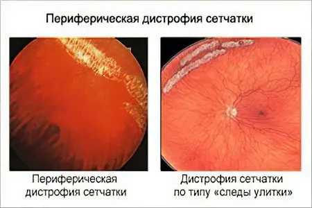
Dystrophy, the damage of which proceeds according to the type of snail track. On examination, whitish, somewhat gleaming perforated defects are visible, it is because of this that this type of disease received its name. At the same time, they merge into ribbons and resemble the trace of a snail. Often large gaps form as a result of this type of dystrophy. In most cases, it is observed in people with myopic disease, it is less common than lattice dystrophy.
Hoarfrost dystrophy is inherited, the changes are bilateral and symmetrical. This type of dystrophy got its name due to the fact that inclusions are formed on the retina, resembling snow flakes, somewhat protruding above its surface.
Dystrophy by the type of cobblestone pavement characterized by the formation of deeply located white annular defects having an oblong shape. Their surface is even, in 205 cases it is observed in patients with myopia.
Retinoschisis – in most cases, this defect is hereditary and is characterized by retinal detachment. Sometimes it occurs in myopia and in old age.
Small cysts dystrophy – characterized by the formation of cysts that have the ability to merge, their color is red, the shape is round. When they break, perforated defects are formed.
Symptoms of peripheral retinal dystrophy
Regardless of the type of peripheral dystrophy, patients complain of similar symptoms:
Visual impairment. Sometimes it occurs in only one eye, sometimes in both.
Restriction of the field of view.
The presence of fog before the eyes.
Distorted color perception.
Rapid fatigue of the organ of vision.
The presence of flies or bright flashes before the eyes. This symptom is intermittent.
Image distortion, the picture looks like a person is trying to see through a thick layer of water.
Violation of the perception of the shape of a real object and its color – metamorphopsia.
Decreased vision in poor light or at dusk.
Symptoms can occur both in combination and separately. They get worse as the disease progresses. The danger of peripheral dystrophy lies in the fact that at the initial stages the disease does not manifest itself in any way, but develops asymptomatically. The first signs may begin to disturb a person when, for a year, the detachment reaches the central sections.
Causes of peripheral retinal dystrophy

Among the causes of peripheral dystrophy are the following:
The hereditary factor, it has been proven that dystrophy occurs more often in those people whose loved ones suffered from a similar problem.
Myopia, this is due to the fact that the length of the eye increases, its membranes stretch and become thinner at the periphery.
Inflammatory eye diseases.
Eye injuries of various origins, including craniocerebral.
High blood pressure.
The presence of atherosclerosis in history.
Diabetes.
Infectious diseases.
Carrying weights, diving under water, climbing to heights, any extreme physical exertion on the body.
Intoxication of the body.
Chronic illnesses.
The disease does not depend on age and gender, it occurs with the same frequency in men, women, children and pensioners.
Treatment of peripheral retinal dystrophy
Before proceeding with the treatment of peripheral dystrophy, it is necessary to correctly diagnose it. The difficulty lies in the fact that the symptoms at the initial stage of the development of the disease practically do not manifest themselves in any way, and during a routine examination, the periphery zone is inaccessible to an ophthalmologist. Therefore, a thorough and systematic examination is necessary, in the presence of risk factors.
Laser coagulation. Treatment of peripheral retinal dystrophy primarily consists of surgery. To do this, use the method of laser coagulation of blood vessels, which consists in delimiting the zone damaged by dystrophy. Laser coagulation can also be carried out for prophylactic purposes in order to prevent the development of dystrophy. This is not a traumatic operation that does not require a rehabilitation stage and a person in a hospital. After it is completed, it is advisable for the patient to prescribe medication and physiotherapy courses.
For preventive measures refers, first of all, to regular visits to an ophthalmologist, especially by people at risk. Peripheral dystrophy is dangerous for its complications, which is why its early diagnosis and timely treatment are so important. It is important to remember that treating peripheral dystrophy is laborious, but preventing the disease is much easier. Therefore, a preventive visit to an ophthalmologist is so important, because any, even modern treatment, is not able to restore vision by 100%, and the goal of surgical intervention will be to stop the gaps and stabilize the level of vision that a person has for the period of treatment.
Author of the article: Degtyareva Marina Vitalievna, ophthalmologist, ophthalmologist


