Contents
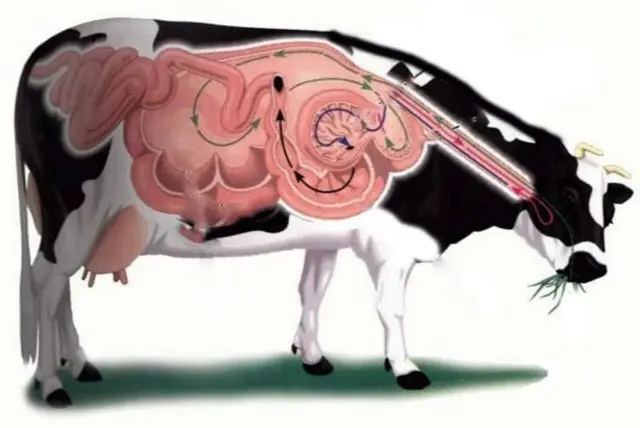
Paramphistomatosis in cattle is a disease caused by trematodes of the paramphistomat suborder, which parasitize in the gastrointestinal tract of cows: the abomasum, scar, net, and also in the small intestine. Infection with paramphistomatosis occurs by the alimentary route when animals graze in the area of flood meadows, in floodplains of rivers with water and grass. The acute course of the disease begins a few weeks after the parasite enters the body of cattle.
Pathology causes significant damage to cattle breeding along with other parasitic diseases of cows. The disease is widespread in Australia, Europe, Asia and Africa. Cases of paramphistomatosis of cattle are constantly recorded in Ukraine and Belarus. On the territory of Our Country, it occurs in different seasons in some areas of the Central Region, in the Black Earth Region, in the Far East and in the south of the country.
What is paramphistomatosis
Cattle paramphistomatosis is a helminthic disease. It is characterized by an acute and chronic course with a lag in the development of animals, and in young individuals there is a high probability of death.
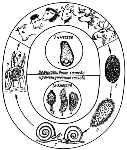
The causative agent of the disease in cattle is a trematode. It is small in size – up to 20 mm. It has a spindle-shaped body of a pink hue. Round in cross section. It is fixed with the help of the ventral sucker at the posterior end of the body, while the oral sucker is absent. From the reproductive organs there is a testicle, uterus, zheltochnik, ovary. Intermediate hosts for them are various types of molluscs.
Helminth eggs are quite large, rounded, gray in color. Excreted into the environment with animal feces. At a comfortable temperature for them (19-28 ° C), after a couple of weeks, meracidium (larva) emerges from the eggs. It penetrates into the body of the shell mollusk, forming maternal redia in its liver. After 10-12 days, daughter redia are formed from them, in which the development of cercariae occurs. They remain in the body of the intermediate host for up to 3 months. Then they go outside, attach to the grass and become infectious for cattle. After being swallowed by animals, adolexaria are released from the cysts and penetrate into the mucous membranes, attaching to the villi.
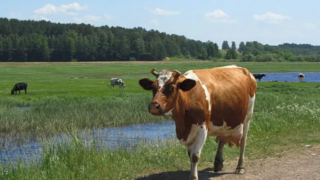
Cattle can become infected with paramphistomatosis in a pasture during a watering place. Paramphistomats are localized in the intestinal mucosa of the individual and move into the rumen. There is a period of puberty, which lasts about 4 months.
Symptoms of paramphistomatosis in cattle
Clinical symptoms are most pronounced in the acute course of paramphistomatosis. KRS notes:
- oppression, general weakness;
- lack of appetite;
- indomitable thirst;
- development of anorexia;
- diarrhea with an admixture of blood and mucus that does not stop for more than a month;
- a dull, disheveled coat and sunken flanks are noted;
- fever;
- rapid depletion of the body;
- tail, hair in the anus area stained with feces.
The chronic course of paramphistomatosis in cattle is more often the result of an acute illness or the gradual spread of parasites by young individuals over a long period of time by a small number of trematodes. At the same time, cattle suffer from prolonged incessant diarrhea, anemia, swelling of the dewlap and intermaxillary space, and a decrease in fatness. Dairy cows dramatically lose productivity.
Sexually mature individuals of paramphistomats more often affect the organism of infected livestock locally. While young trematodes, parasitizing in the intestines and abomasum, cause their significant changes. Therefore, the disease in young cattle is difficult and often ends in the death of animals. Paramphistomatosis is exacerbated by secondary infection as a result of mechanical and trophic action.
Diagnosis paramphistomatosis
The diagnosis of paramphistomatosis of a diseased individual of cattle is made, taking into account epizootological data, clinical manifestations of the disease and laboratory tests.
The acute form of paramphistomatosis is diagnosed by fecal helminthoscopy. To do this, 200 g of feces are taken from cattle for analysis and examined by successive flushing. The efficiency of this method is about 80%. Helmintocoproscopic studies are carried out to identify the chronic form of the disease. Cattle paramphistomatosis, a particularly acute manifestation of the disease, should be differentiated from a number of other similar pathologies.
Dead animals are autopsied. Carefully examine the stomach, duodenum, abomasum, scar. The veterinarian notes the general exhaustion of the cattle that died from paramphistomatosis, gelatinous infiltrate in the intermaxillary space, swelling and hemorrhagic inflammation of the duodenum and stomach. The gallbladder is greatly enlarged, contains mucus and trematodes. Young parasites are often found in the abomasum, bile ducts, peritoneum, and renal pelvis. Traces of blood are visible in the small intestine of cattle. Lymph nodes with paramphistomatosis are edematous and somewhat enlarged.
Treatment of paramphistomatosis in cattle
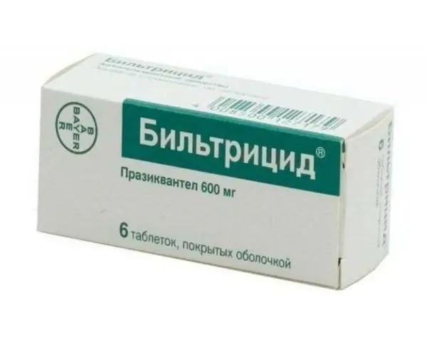
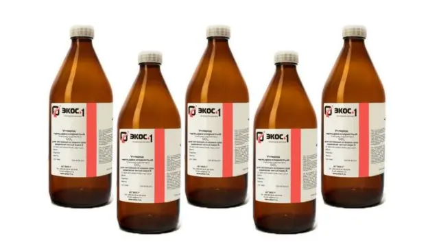
Veterinary specialists consider the drug Bitionol or its analogue biltricid to be the most effective remedy against ruminant paramphistomatosis. It is prescribed to cattle in a dosage depending on the body weight of the diseased animal after a starvation diet for 12 hours. It should be applied twice with an interval of 10 days. Based on the condition of the individual, symptomatic treatment is carried out.
Prevention of paramphistomatosis in cattle
Farms suffer huge economic damage when paramphistomatosis occurs in cattle. The main preventive measures should be aimed at preventing the disease, since it is quite difficult to deal with it and sometimes it is impossible to achieve a full recovery.
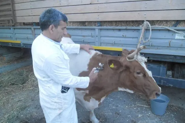
Cattle breeders should not let young cattle go for walking, it is better to make a separate paddock for them, create an artificial upland pasture away from various water bodies. It is necessary to carry out deworming in a timely manner before the start of the stall period with laboratory control by veterinarians. Flooded pastures should be examined for the presence of an intermediate host – shell mollusks. When it is detected, grass from these places should not be fed to animals. First, pastures are drained, plowed, checked again, then used for their intended purpose. Watering cattle during grazing is possible only with imported water. Manure should be biothermally disinfected.
Conclusion
Paramphistomatosis in cattle is a disease that is extremely difficult to get rid of. Often it leads to the death of animals and infection of the entire herd. Paramphistomatosis causes serious damage to farms. Sometimes up to 50% of the cattle population die from it, the productivity of dairy cows decreases. At the same time, preventive measures are quite simple, one of which is deworming the herd.


