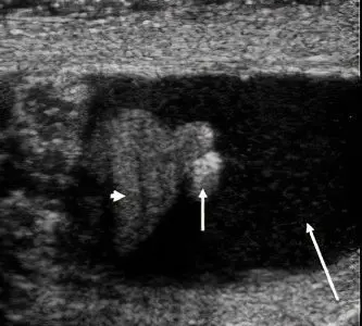
Hydatida Morgagni is a rudiment in boys and men; in women, internal genital organs are formed from it. Hydatida Morgagna is a pedunculated polyp that can be located on the testicle, epididymis, appendix, or vas deferens. The hydatids of the testis and epididymis were described in 1761 and named after the author. The testicular process is located in the area of connection with the head of the epididymis, is designated as the embryonic remnant of the Müllerian duct.
And the process of the appendage located on its head is a rudimentary part of the cranial part of the Wolffian duct. The aberrant ducts of Haller act as hydatids in the region of the body and tail of the appendage, and paradidymis in the region of the distal part of the spermatic cord. Almost all hydatids have a stalked structure, some are characterized by a wide base, with a diameter of 0,2 to 1,5 cm.
Morgagni’s hydatids contain cystic inclusions, are covered with cylindrical epithelium, consist of connective tissue formations, with delicate and loose stroma, thin and fragile blood vessels. These embryonic formations are easily exposed to various pathologies, among which there is necrosis provoked by injuries – jumping from a height, outdoor games, scrotal trauma and many more reasons. Hydatid necrosis Morgagni most often occurs as a result of its torsion, which disrupts the work of blood vessels supplying oxygen.
This is necessarily accompanied by the development of the inflammatory process, hemorrhage and necrosis. Necrosis develops during infectious processes or after minor injuries. Clinically, Morgagni’s hydatid necrosis is manifested by edema, inflammation, and impaired blood flow throughout the scrotum, which often leads to testicular degeneration. Hydatid necrosis develops rapidly, therefore it refers to emergency pathologies.
By opening the membranes, an accurate diagnosis is made, its fidelity is confirmed by a cloudy, purulent, effusion with fibrin flakes.
But it is not always possible to accurately diagnose it, it can be confused with orchitis, epididymitis, etc. The symptoms of the disease are constant, gradually increasing pain in the groin, lower abdomen, in the testicle area. Visually, you can see a dark blue knot translucent through the tissue of the scrotum. Palpation due to the presence of dropsy and severe pain is impossible. In the initial period of the development of changes, a diagnosis can be made on the basis of external signs. .
Usually, on the 2nd day, the testicle is affected by dropsy, swelling and redness of the scrotum appear. Hyperemia of the scrotum and secondary dropsy (hydrocele) are late signs of the disease. Hydatid necrosis causes a condition called edematous scrotum syndrome, which has nuances in its development and reflects a picture of underlying disorders. If, after elimination of torsion by warming or blockade with novocaine of the spermatic cord, the dark color characteristic of necrosis does not change, then the testicle is removed.
A timely operation with a pronounced syndrome of edematous scrotum contributes to the preservation of the organ, otherwise testicular atrophy may develop. Torsion-related necrosis may occur in utero, after birth there is a painless hard mass in the scrotum, the color of the scrotum changes, signs of ecchymosis and edema may be detected. The viability of the testicle in this case is impaired. In order to prevent the phenomenon of torsion and necrosis in the future, fixation of a second, healthy testicle should be performed.
Hydatid necrosis can occur in any inflammatory process, is a consequence of erysipelas of the skin, weeping eczema. At the same time, ischemic areas are formed in the tissues of the suspension, and aseptic chronic vaginitis develops. The disease is characterized by the severity of the course in the presence of anaerobic infection. In the advanced stage, blisters with serous-hemorrhagic contents appear on the skin, patients complain of headache, high body temperature, chills, shortness of breath, and palpitations.
When bubbles open spontaneously, erosions form. The appearance of dark gray spots indicates necrosis of the scrotum. A week later, the formed demarcation line contributes to the rejection of dead skin of the scrotum and the release of pus with gas bubbles. The process of melting and rejection can last up to 12 days from the onset of the disease. When the testicles are completely exposed, pain disappears, the temperature decreases and a period of skin regeneration begins.
For the diagnosis of acute diseases of the organs, thermography, radioisotope scanning, ultrasound echotomography, puncture of the scrotum and testicles with a thick needle are used. Ultrasound studies can accurately determine the degree of functional and structural changes.


