


Contents
In line with its mission, the Editorial Board of MedTvoiLokony makes every effort to provide reliable medical content supported by the latest scientific knowledge. The additional flag “Checked Content” indicates that the article has been reviewed by or written directly by a physician. This two-step verification: a medical journalist and a doctor allows us to provide the highest quality content in line with current medical knowledge.
Our commitment in this area has been appreciated, among others, by by the Association of Journalists for Health, which awarded the Editorial Board of MedTvoiLokony with the honorary title of the Great Educator.
Neck ultrasound is an imaging test that is used to assess the condition of organs in the neck and mouth. Ultrasound can target the thyroid gland, larynx, lymph nodes or salivary glands. The test is recommended when the patient has pain in the neck and adjacent organs, or the doctor suspects specific diseases of these areas.
Neck ultrasound – a safe imaging test
Neck ultrasound is a painless and non-invasive method of imaging structures and organs in this area of the body. The doctor uses equipment that emits harmless ultrasonic waves. They propagate in the examined medium and reflect on its border to return to the receiver. The computer processes the data obtained, and the finished image can be viewed on the monitor.
Depending on the structure studied ultrasound device is set to the appropriate frequency. This allows the test parameters to be adjusted to different tissues and organs, as they may differ in the degree of absorption of ultrasound waves. Neck ultrasound is used to diagnose lymph nodes, soft tissues (tendons, muscles), larynx, as well as the thyroid gland, parathyroid glands and salivary glands.
Neck ultrasound – indications for examination
Execution ultrasound examination of the neck it is recommended in the case of pain, swelling or palpable enlargement of organs (larynx or thyroid gland). Often, imaging of the cervical structures is done with a focus on the thyroid gland. The ultrasound of this organ is performed when the thyroid gland is enlarged or the results of hormonal tests (THS, fT3, fT4) are incorrect.
Other indications for a neck ultrasound include:
- enlargement or soreness of the cervical lymph nodes;
- sore throat or mouth which gets worse when swallowing;
- enlargement of the parotid and submandibular glands;
- pain in the salivary glands under pressure;
- pains radiating to the ears;
- control of the condition of the thyroid gland in people with an overactive or underactive thyroid gland.
If there is a suspicion of circulatory abnormalities, Doppler ultrasound of the carotid and vertebral arteries can be performed. The diagnosis of specific pathological changes (nodules, thickenings) in the neck area is possible thanks to ultrasound-guided fine-needle biopsy.
Read more about:
- Ultrasound of the thyroid gland and parathyroid glands;
- Ultrasound of the salivary glands.
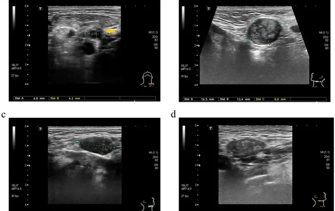
What does an ultrasound of the neck detect?
On the basis of ultrasound examination of the neck, inflammatory, purulent and neoplastic changes are diagnosed. Due to the easy access, speed of execution and the lack of contraindications, ultrasound is the first imaging examination recommended for suspected pathology of the cervical structures.
Thyroid ultrasound results may indicate, among others on:
- inflammation – enlarged lymph nodes or abscesses;
- neck phlegmon;
- congenital cysts;
- ductal urolithiasis of the salivary glands;
- tuberculosis of the lymph nodes;
- actinomycosis;
- cat scratch disease;
- benign and malignant tumors of neoplastic etiology, including lipomas, non-Hodgkin’s lymphomas, Hodgkin’s lymphoma, sarcomas.
Contraindications for neck ultrasound
Neck ultrasound is recommended to practically everyone, because there are no contraindications that would completely exclude the possibility of implementing the procedure. The only situations that are a premise for abstain from testing are skin burns and open wounds. When one of the symptoms of the examined person is pain in the examined area, the pressure of the head used during the ultrasound of the neck may cause discomfort.
Neck ultrasound – how to prepare?
In most cases, you do not need to prepare for neck ultrasound. It is recommended to choose comfortable clothing, as the neck must be exposed during the examination. Another point to think about before visiting relates to men. The facial hair should be shaved off before the examination, because it can make it difficult to perform – the ultrasound gel is spread directly on the skin, and the head should be in direct contact with its surface.
What does an ultrasound of the neck look like?
You should take a referral on ultrasound of the neck, if you have received any. We will perform the examination under the NFZ contract, but we can also attend it privately. In most cases, the waiting time for an ultrasound of the neck is similar, from several days to a week.
Neck ultrasound price depends on the scope of the study. If we examine only the lymph nodes or salivary glands, we will pay from 60 to 150 PLN. A wider assessment of the neck and craniofacial organs (including the thyroid gland, larynx, salivary glands, lymph nodes and soft tissues) is associated with a higher cost, reaching approximately PLN 150-250. Another variation of the procedure is the ultrasound Doppler of the carotid and vertebral arteries, the price of which starts around PLN 120.
- Also read: Doppler ultrasound – what is it, when to perform it and what it diagnoses
Duration of ultrasound of the neck as a rule, it does not exceed 20 minutes. The first stage is a short medical interview, during which we present the problem and provide the results of previous imaging tests, if any. Then we lie down on the couch and reveal the neck. The doctor covers the skin with gel and then places an ultrasound probe on it. He observes the image obtained on the monitor, it is also saved on a memory device, and if necessary, a specific shot is also saved in the form of a printout.
After the test is completed, a description of the results is prepared. With the received documentation, go to the doctor who referred us for the examination. He will assess the cervical structures and advise on further suggested procedures.