Monocytes are necessary for the release of the body from foreign microorganisms and toxic substances. The processes of capturing, blocking and destroying pathogens occur with virtually no negative consequences for these most massive blood cells. After interacting with uninvited guests, monocytes retain their properties and continue their journey through the bloodstream.
Among the total volume of leukocyte blood elements, monocytes (MON) belong to 2 to 10% of the cells. Medical terminology under the definition of “monocytes” means mononuclear phagocytes, histiocytes and macrophages. The pronounced bactericidal properties of blood elements are fully manifested in an acidic environment. A change in the number of monocytes, both up and down, signals pathological changes in the body, possibly even the onset of a serious illness.

What are monocytes, the mechanism of their formation
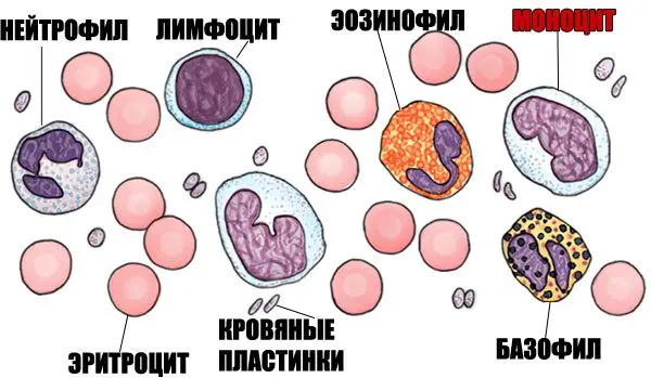
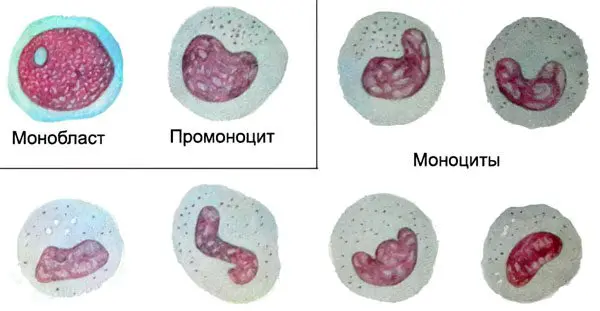
Monocytes – white blood cells with a diameter of 18 to 20 microns, related to agranulocytic leukocytes. These are the largest cells in the peripheral circulation. Microscopically, a monocyte is defined as an oval cell with a single polymorphic nucleus resembling a bean in shape. The nucleus occupies an eccentric position. The pronounced staining of the monocyte nucleus distinguishes it from lymphocytes. When evaluating a blood test, this sign allows you to correctly assess the blood picture. Healthy indicators of monocytes are 3-11% of the total volume of white blood cells.
A significant number of these cells is determined in:
spleen;
liver;
lymph nodes;
bone marrow.
The formation, development and growth of monocytes proceeds to the bone marrow under the influence of active substances:
Glucocorticosteroids that can slow down the synthesis of monocytes.
Cell growth factors GM-CSF and M-CSF, which stimulate the development of monocytes.
Formed mature monocytes function in the bloodstream for 2-3 days. Some of them die during this period due to physiological apoptosis, provided for by normal natural processes in the body. The remaining cells transform into macrophages and spread throughout the internal organs and tissues. The life cycle of these advanced cells is 1 to 2 months.
Special qualities of monocytes
The main part of monocytes is synthesized in the bone marrow. The first to emerge from a multipotent stem cell is a monoblast. In the future, it goes through the promyelomonocyte phase, and then the promonocyte phase. Promonocytes in the last stage of maturation have a pale friable nucleus, traces of nucleolus are visible in the cytoplasm. Azurophilic granules are determined in the composition of mature monocytes and promonocytes. Both types of cells are of the agranulocytic type. Granules of immature cells, histogenic particles and lymphocytes are the result of cytoplasmic protein discolloidosis and are stained with azure according to the Romanovsky-Giemsa method. In the connective tissue of internal organs and lymph nodes, an insignificant part of monocytes is formed.

In the cytoplasm of mature monocytes, biologically active substances, hydrolytic enzymes were found:
Lipase.
Protease.
Verdoperoxidase.
Carbohydrase.
Lactoferrin and myeloperoxidase are found in minimal amounts.
The human body is able to activate the synthesis of monocytes in the bone marrow slightly. The division of phagocytic mononuclear cells outside the bone marrow is extremely slow and limited. The mechanism of replacing dead specific cells is possible only with the help of monocytes that spread through the bloodstream. In the peripheral blood, monocytes function for about 72 hours, after which they penetrate into the tissues of nearby organs. Here the process of their transformation into mature histocytes, highly differentiated macrophages – Kupffer cells of the liver, alveolar cells of the lungs and other organs is completed.


