


Contents
In line with its mission, the Editorial Board of MedTvoiLokony makes every effort to provide reliable medical content supported by the latest scientific knowledge. The additional flag “Checked Content” indicates that the article has been reviewed by or written directly by a physician. This two-step verification: a medical journalist and a doctor allows us to provide the highest quality content in line with current medical knowledge.
Our commitment in this area has been appreciated, among others, by by the Association of Journalists for Health, which awarded the Editorial Board of MedTvoiLokony with the honorary title of the Great Educator.
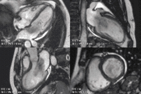
Magnetic resonance imaging is one of the most accurate imaging tests used in cardiological diagnostics. It is recommended when the results of basic tests raise doubts or when other diagnostic forms are difficult. Magnetic resonance imaging of the heart is safe, and it can be performed with enhancement in the form of a contrast agent. What conditions will MRI of the heart detect?
Magnetic resonance imaging of the heart – when to perform?
Cardiac imaging diagnostics is recommended for people suffering from heart diseases or after a heart attack. It is also performed when your doctor detects heart murmurs or arrhythmias during basic checkups. An additional indication of the patient’s symptoms, including pain, shortness of breath, palpitations.
The patient is first referred to a chest X-ray or echocardiography, commonly known as the echo of the heart. If these tests are difficult to perform or if there are clinical uncertainties, cardiac magnetic resonance imaging may be the solution. This is a high-precision imaging test. It enables the assessment of the work of the heart, blood flow, correct valve closure or changes in the pericardium.
Thanks to magnetic resonance imaging of the heart, it is possible to detect:
- congenital and acquired heart defects;
- cardiomyopathy;
- arrhythmia;
- myocardial infarction;
- coronary artery disease;
- valvular defects;
- tumors;
- thrombus;
- scarring changes and fibrosis;
- pericardial diseases.
Magnetic resonance imaging of the heart allows for a detailed analysis of the anatomy of the heart and its functioning. On this basis, it is possible to accurately diagnose the causes of dysfunction or pathological changes, e.g. inflammation or ischemia.
In the diagnosis of heart diseases, computed tomography, coronography and scintigraphy are also used.
How does magnetic resonance imaging work?
Magnetic resonance imaging is carried out in a special room where we will find an MRI scanner. The patient lies down on a retractable table, which is set in such a position that the examined part of the body is within the reach of the tube. When the apparatus is turned on, a strong magnetic field is generated, which affects the hydrogen atoms present in the human body, causing them to be organized.
Then, radio waves (RF) are emitted, which meet said atoms, and then, thanks to the resonance phenomenon, return to the computer, which can transform the information obtained into an image.
The examination is safe, it has no side effects and you do not need to limit the number of examinations, as is the case with x-rays. However, the magnetic field can be hazardous when there are metal foreign bodies and electronic devices in the patient’s body.
Are you looking for an MRI facility? You can find it in the clinics.pl database.
- Read more: Magnetic resonance imaging – when is it worth doing and how to prepare for it?
When is an MRI of the heart not performed?
Even for such a non-invasive examination as magnetic resonance imaging, there are contraindications. The patient is disqualified from cardiac MRI with a pacemaker, a cochlear implant, a central nervous system stimulator, some metal intracranial clips, and an insulin pump.
Relative contraindications to inform your doctor include joint prostheses, foreign bodies in the body (e.g. metal filings), sutures, braces and the first trimester of pregnancy.
- Also, check what is full body magnetic resonance imaging.
Preparation for MRI of the heart
Before performing magnetic resonance imaging of the heart, one should remember not only about contraindications and the necessity to leave certain items outside the room with the MR apparatus.
- It is important not to disturb the heart with anything, as this could falsify the MRI result. This means that on the day of the examination, physical activity, drinking coffee or smoking are not recommended.
- People come for the examination on an empty stomach.
- Before the examination, it is important to empty the bladder, as the procedure usually takes several dozen minutes.
- Unless otherwise instructed by the doctor, we normally take medications for chronic diseases.
- In the case of MRI with contrast, a cannula is inserted before the examination. The possibility of providing contrast is verified by blood creatinine tests, which are performed several days or up to two weeks before the MRI.
- During the examination, the patient should not show signs of infection. If he has been taking antibiotics, an MRI is done two weeks after stopping them.
You should also remember not to bring metal objects such as coins, jewelry, glasses, keys into the examination room. This also applies to clothing with metal zippers, buttons or clasps. The magnetic field generated by the MRI machine will also damage magnetic cards, hearing aids and mobile phones.
MRI of the heart – what else is worth knowing?
Below, some additional information on MRI in cardiac diagnosis.
- In the case of young children and people with claustrophobia, it is possible to perform a cardiac MRI under general anesthesia.
- The price of an MRI of the heart oscillates around PLN 600.
- The MRI of the heart takes 30 to 60 minutes.
- During the MRI, the patient receives instructions from the person conducting the examination. One recommendation might be to hold your breath for a certain period of time.
- You have to wait a minimum of a few days for the results of the MRI. During this time, a description is prepared, which is an integral part of the results.