Contents
Lung cancer is by no means a rare disease. Against the background of an aggressive environment, smoking, and previous diseases of the respiratory organs, cancer is widespread. In the initial stages, this pathology is difficult to identify.
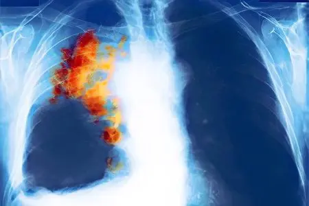
An x-ray of the lungs with cancer can help oncologists in determining the presence of a tumor.
Symptoms indicating a disease

Cancer is an insidious disease that can masquerade as other diseases. But if suspicions of lung cancer are confirmed by such signs as:
Hoarse or wheezing breath;
Lack of appetite;
Bouts of prolonged coughing;
shortness of breath;
Increased body temperature;
Lethargy, apathy, then you need to seriously take up the clarification of the clinical picture of the neoplasm of the lungs.
How is lung cancer diagnosed today?
Modern diagnostics of lung oncology has in its arsenal four categories of examination for neoplasms:
First. Methods confirming the possibility of a tumor development process. This is a medical examination, a fluorographic study, an x-ray for lung cancer.
Second. The diagnosis is confirmed on computed tomograms. A bronchoscopic and radionuclide examination is also carried out.
Third. This includes methods of the morphological plan. In this case, the diagnosis of a lung tumor is decisively determined. Histology and cytology of tumor specimens is done using endoscopy or biopsy.
The last, fourth category of diagnostics makes it possible to identify the degree of prevalence of the oncological process. This requires ultrasound, CT and radionuclide research.
What results does an x-ray show?
The information content of such a study allows you to determine lung cancer on an x-ray. It happens that only a few cases of a similar clinical picture show a satisfactory condition of the lungs.
An x-ray image will help to detect the central form of cancer by darkened areas of the lungs and an expanded network of pulmonary vessels.
On a frontal x-ray, the tumor looks like a clear, even shadow from the edge of the lungs to the right / left side. Ribbon-shaped processes extend from this shadow.
A photo of what lung cancer looks like on an X-ray examination is not surprising for oncologists, but the patient is of little pleasure.
Is lung cancer visible on fluorography
Fluorography is the same study as x-rays, it consists in photographing the respiratory organs on a fluorescent screen. X-rays pass through the body, and as a result of the uneven absorption of X-rays by different tissues of the body, an image is obtained that specialists can decipher.
Fluorography gives a small size of the organ under study, but for oncologists this is enough to make a diagnosis from an x-ray photo.
Bronchogenic cancer
Under this name, lung cancer is best known among physicians. Why bronchogenic? Yes, because this pathology arises from the bronchial mucosa. The walls of the bronchi are the most vulnerable part of the internal organs, because only they have direct contact with the environment and the toxic substances in it.
As a result, with the help of inhaled carcinogens, the tumor grows intensively. And it is growing by leaps and bounds.
Therefore, the question: “Does the fluorogram show lung cancer” can be answered in the affirmative – it does. This diagnostic method is convincing, it can show the disease, as well as determine at what stage it is.


