Contents

HSG X-ray (X-ray hysterosalpingography, WG-HSG) is a diagnostic technique designed to assess the condition of the female genital organs. The essence of the study is that a catheter is inserted into the uterine cavity, through which a contrast agent is supplied. It is this that will be seen during the performance of a series of X-ray images. After it is evenly distributed throughout the uterus and appendages, the doctor “sees through” the organs using an X-ray machine. The resulting images will clearly show the fallopian tubes and uterus.
Using this procedure, it is possible to detect such pathologies of the genital organs as obstruction of the fallopian tubes, endometriosis growths in the uterus, anomalies in the structure, etc. This method is often recommended for women who suffer from infertility, since these factors most often lead to the fact that patients cannot get pregnant.
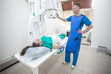
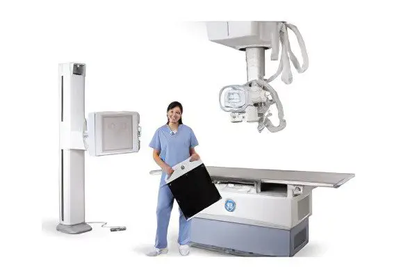
It is possible to perform the procedure both in an outpatient clinic and in hospitals of gynecological departments. The main condition for its implementation is the presence of an X-ray apparatus and a specialist who knows how to work on it.
During hysterosalpingography, the woman is in the gynecological chair. After the doctor injects a contrast agent into the uterine cavity, it is necessary to withstand some time. This will allow the liquid to be evenly distributed throughout the internal genital organs. After that, one or more pictures are taken, which allow you to evaluate the result.
If a woman does not have any pathologies, then the uterus looks like a regular triangle, and the tubes are arched. In the presence of diseases, the picture changes: it is possible to detect neoplasms (polyps, myomas), partitions, adhesions, etc. If a woman has obstruction of the fallopian tubes, then the contrast agent will be distributed unevenly, and obstacles in its path will be clearly visualized.
The procedure does not require the introduction of anesthesia, as it does not cause pain. However, if a woman has a high threshold of pain sensitivity, then local anesthesia is indicated for her.
HSG and ultrasound – is it the same thing?

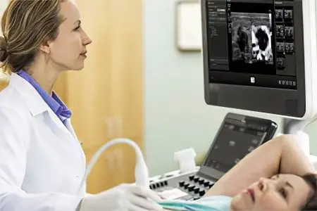
HSG and ultrasound are two different procedures. Ultrasound examination involves an overview of the internal organs of the patient and the detection of a possible pathology by changing their structure and density. The picture is displayed on the monitor. To perform an ultrasound, there is no need to perform any additional procedures. It is enough just to lubricate the surface to be viewed with a special gel.
HSG involves the introduction of a contrast fluid into the uterus. After its distribution, the doctor performs a series of images using an X-ray machine (but it is possible to examine the internal organs on an ultrasound machine). The introduction of a contrast agent allows you to make the study more informative. In addition, the doctor can diagnose blockage of the fallopian tubes, which cannot be done during a conventional ultrasound examination.
Since two devices can be used for HSG: X-ray and ultrasound, there is a difference in the course of diagnostics. If the pictures are taken on x-ray equipment, then the procedure is called “X-ray hysterosalpingography”. When an ultrasound machine is used to perform the study, the technique is called echohysterosalpingography. Due to the similarity in name, many people believe that these procedures are identical, in fact, their essence and diagnostic significance are different.
Which is better: X-ray or HSG?

A standard x-ray examination will not reveal obstruction of the fallopian tubes or other pathologies of the pelvic organs, so it is never used for this purpose. HSG, on the contrary, is the method of choice for suspected obstruction of the fallopian tubes, endometrial polyps, uterine myoma, endometriosis and other pathologies of the female reproductive system. Therefore, HSG is definitely better than X-ray.
However, it should be understood that HSG is performed either with the help of an X-ray machine or with the help of an ultrasound diagnostic machine. The internal genital organs of a woman become visible on them after the introduction of a special contrast agent into the uterus and fallopian tubes. Many specialists use the term “X-ray” to refer to the procedure for examining the fallopian tubes on an X-ray machine, and the term “HSG” to conduct a study on an ultrasound machine. If we consider the issue from this point of view, then the GHA will be better than “X-ray”.
The fact is that Echo-HSG has the following advantages over RGHA:
A woman will not have to use contraceptive methods that protect her from pregnancy if she has been prescribed an ultrasound HSG.
There are no contraindications for conceiving a child in the month when Echo-HSG was performed.
During the procedure and after it, there is no risk of an allergic reaction to a contrast agent that contains iodine.
The patient’s body will not be irradiated by the x-ray machine. Moreover, this negatively affects the number of eggs that are present in the ovarian follicles (ovarian reserve).
Indications for HSG

HSG of the fallopian tubes is carried out by gynecologists and gynecologists-oncologists.
The indications for the procedure are as follows:
tubal infertility;
Adhesions of the pelvic organs;
Anomalies in the development of the reproductive organs;
Sexual infantilism;
Fibroma of the uterus, located in its submucosal layer;
endometrial cancer;
Endometrial growths in the uterine cavity;
Polyps;
hyperplasia of the endometrium.
Hysterosalpingography is the method that will confirm that a woman has a pathology of the fallopian tubes or uterus, but does not always make it possible to assess the severity of the disease and its nature. If we turn to statistics, then in 98% of cases it is possible to identify an existing violation, but it is possible to make a correct diagnosis only in 35% of cases.
Contraindications for HSG

X-ray hysterosalpingography may not always be performed.
There are certain contraindications to the procedure, including:
Intolerance to iodine preparations. This contraindication is due to the fact that the composition of the contrast agent, which is injected into the uterine cavity, contains iodine.
Inflammation of the ovaries, uterus, appendages.
Colpitis, cervicitis, bartholinitis.
Acute infectious diseases of the body, for example, acute respiratory infections, acute respiratory viral infections, influenza, tonsillitis, sinusitis, etc.
Violation of blood clotting.
Heart disease.
Renal failure.
Severe disorders in the liver.
Pathology of the thyroid gland.
Pregnancy. Carrying out a pregnancy test is a prerequisite before undergoing the procedure.
Menstrual bleeding.
Lactation.
Increased ESR and leukocytosis.
Preparing for the procedure

Preparation for the procedure is simple, but following the recommendations given by the doctor is a prerequisite. Otherwise, you can harm your own body.
So, a woman must follow the following rules in order to qualitatively prepare for hysterosalpingography:
1-2 days before the proposed study, you need to give up intimacy.
7 days before the procedure, do not douche or use intimate hygiene products inserted into the vagina.
7 days before the study, it is forbidden to use vaginal tablets, suppositories and ointments for treatment.
2-3 days before the study, you need to change your diet, refusing to eat foods that provoke excessive gas formation. This applies to cabbage, legumes, bread, dairy drinks, soda.
7 days before the procedure, you should stop using tampons.
After the end of the next menstruation, partners must use a condom to avoid conception.
It is equally important to undergo a qualitative examination before the GHS. It necessarily includes the delivery of the following tests:
Oak
OAM.
TANK.
Blood test for syphilis, HIV, hepatitis.
A smear from the vagina and from the cervix.
Before you go for the procedure, you need to remove all hair from the external genital organs, wash them thoroughly. The bladder and bowels must be empty. If it was not possible to go to the toilet, then an enema should be done. The procedure must be carried out on an empty stomach.
As for self-administration of painkillers, it is forbidden to use medicines without consulting a doctor. As prescribed by the doctor, it will be possible to drink an antispasmodic, for example, No-shpu, 30 minutes before the HSG.
When is the HSG performed?
Most often, hysterosalpingography is performed within 2 weeks after the next menstruation. This is due to the fact that during this period the uterine mucosa has a small thickness, which means it does not block the entrance to the fallopian tubes.
Although, depending on the purpose of the study, it may be scheduled at other times. To assess the patency of the fallopian tubes, it is carried out in the second half of the menstrual cycle. If there is a suspicion of internal endometriosis, then it is recommended to perform HGS on the 7th-8th day of the cycle. Fibroids in the submucosal layer of the uterus can be detected at any time, but only on condition that the woman does not have menstruation.
How is the HSG performed?

If the doctor’s office is equipped with a special X-ray chair, then the woman is seated on it. If there is no such chair, then the patient will be located on a regular gynecological chair, and an x-ray machine will be brought to her.
After treating the external genital organs with an antiseptic composition, the doctor inserts mirrors into the vagina and wipes the vaginal walls first with dry cotton wool, and then moistened with an alcohol solution. The next step is to insert the tube into the cervical canal. The tube is attached with bullet tongs. When this manipulation is done, the mirrors are removed. A contrast agent is supplied through the tube with a syringe, which must first be warmed up to approximately the woman’s body temperature (up to 37 ° C).
When the contrast agent is evenly distributed throughout the uterine cavity and fallopian tubes, the doctor begins to take pictures. As a rule, the doctor takes 4 to 6 pictures during the procedure. To begin with, the state of the uterus (its relief) is fixed. Then another 4 ml of a contrast agent is injected into the cavity, which makes it possible to more clearly visualize the appendages. If this volume of liquid is not enough, then enter as much as necessary.
After all the pictures are taken, the patient is transferred to the couch and left in a horizontal position for another hour. The fluid that was injected during the procedure is absorbed into the bloodstream and excreted from the body by the liver and kidneys.
What contrast agent is used for HSG?

For the procedure, the introduction of a contrast fluid is shown, which has the ability to delay x-rays. These are drugs such as:
CardioTrust. This is a contrast agent that can contain 50% and 30% iodine.
Urotrast, Triombrast and Verographin. These are three analogues belonging to the group of radiopaque substances, which may contain 60% and 76% iodine.
Interestingly, for the first time, hysterosalpingography was tried with Lugol’s solution in 1909. But due to the occurrence of irritation of the peritoneal cavity and uterus, this attempt was unsuccessful. A year later, the Lugol solution was replaced with bismuth paste, and then with argyrol and collargol. However, it was not possible to achieve the desired effect with the use of these substances. In addition, all of them entailed inflammatory processes of the peritoneum.
Only in 1925 did the scientist Heuser first use Lipiodol (a preparation containing iodine) for hysterosalpingography. This substance made it possible to visualize the condition of the uterus and fallopian tubes well, and also did not harm the health of the woman. Since then, the procedure has been introduced into medical practice.
Consequences and complications of HSG

A woman should use sanitary pads for 2-3 days after the procedure. This need is due to the fact that the remains of the contrast agent can flow from the vagina. If a small amount of blood is found in the discharge, then you should not worry about this, since such a phenomenon is a variant of the norm.
Mild pain, reminiscent of those that occur during the next menstrual cycle, should not frighten a woman. After the HSG, such discomfort does not indicate any complications.
Also, the patient should be prepared for the fact that during or several hours after the HSG, a metallic taste in the mouth, dizziness, and palpitations may occur. This is a normal reaction of the body to the introduction of a contrast agent.
A woman should not visit a sauna or a bath, as well as take a hot bath for 3-4 days after undergoing the HSG.
To show concern and immediately consult a doctor should be with an increase in body temperature, heavy bleeding or severe pain in the lower abdomen.
Extensive blood loss, infection, perforation of the uterus and fallopian tubes during the HSG procedure in modern gynecological practice are extremely rare, as an exception.
A woman should refrain from pregnancy for the next 3 months. For protection, you should use a condom.
As a rule, the procedure is well tolerated, but only on condition that the preparation for it is carried out qualitatively: inflammatory processes were detected in time, there are no other contraindications to HSG.
Evaluation of results

The doctor should interpret the results based on the images obtained.
Signs of various diseases on the x-ray picture are as follows:
Noticeable asymmetry of the uterus – the diagnosis of “unicornuate uterus”.
An elongated cervix and a pronounced decrease in the volume of its cavity is the diagnosis of “infantile uterus”.
Short, long, or asymmetric fallopian tubes – the diagnosis is “congenital obstruction of the fallopian tubes.”
The presence of a flask-shaped expansion in the tubes is a possible diagnosis of “fallopian tube adhesions”, or “sactosalpinx”, or a combination of these two diagnoses.
The presence of light areas in the tubes – “adhesions of the fallopian tubes.”
The shape of the fallopian tubes, resembling the shape of smoking pipes, the presence of extensions in them in the form of a flask, a decrease in the volume of the uterine cavity – the diagnosis is “tuberculosis of the genital organs.”
Uneven contours of the uterus, the detection of defects of an oval or other shape – the diagnosis is “uterine polyps” or “endometrial hyperplasia”.
Of course, only the most obvious and common signs of certain diseases were listed. Only the attending physician can make a final diagnosis, evaluating the whole range of results obtained in the course of various studies.
Cons of the GHA
The disadvantages of the procedure are the following points:
A woman receiving a dose of radiation, albeit a small one.
The likelihood of an allergic reaction to the injected contrast agent. Special care should be taken by women with a history of bronchial asthma, as well as allergic patients.
There is a risk of mechanical damage to the epithelial layer of the uterus, which leads to the appearance of bleeding.
Plus GSA
In addition to the fact that X-ray HSG is highly informative, it has another important advantage. The fact is that hysterosalpingography is not only a diagnostic, but also a therapeutic method of influencing the female body. It has been established that approximately 20% of women suffering from infertility successfully become pregnant after undergoing HSG.
Doctors explain this fact by the fact that during the procedure it is possible to improve the patency of the fallopian tubes, since the injected substances “wash” them, eliminating small adhesions.


