Contents

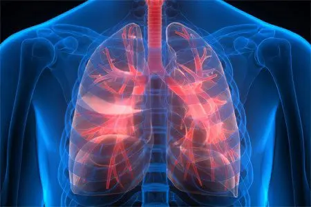
According to medical statistics, the number of patients with lung diseases is increasing every year. Along with the morbidity figures, the mortality rates are continuously increasing. Pneumonia, for example, ranks 4th in the world among the causes of death of the adult population, catching up with cardiovascular and oncological diseases. In figures, this is about 7 million people annually. The main problem is untimely or erroneous diagnosis.
Pneumonia has a second, more common name – pneumonia. The disease affects the alveolar tissue of the lungs with purulent or serous exudate, which leads to the development of a severe clinical picture. The main diagnostic method is X-ray examination. However, it is important to consider that pictures do not always provide complete information. An insufficiently experienced radiologist cannot always see the differences between the signs of pneumonia and lung cancer, tuberculosis.
Lung cancer is often confused with pneumonia. Oncological pathology can disguise itself as other diseases, or generally proceed for a long time without specific symptoms. Tuberculosis is another insidious disease whose early symptoms can be confused with pneumonia. Controversial manifestations of the clinical picture can be differentiated only with the help of special laboratory research methods.
Etiological factors
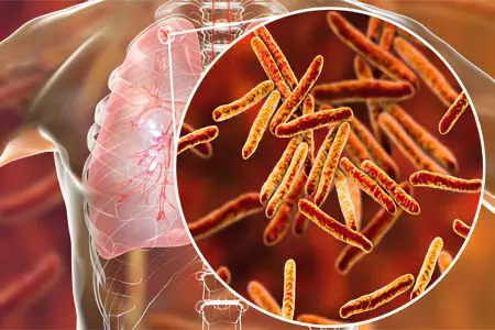
The development of pneumonia is caused by the entry of pathogenic microorganisms into the body through the upper respiratory tract.
The causative agents can be:
Staphylococcus.
Streptococcus.
Haemophilus influenzae.
Klebsiella.
Chlamydia.
Legionella.
Mycoplasma.
In 20%, pneumonia develops during a long recovery period after surgery, injuries that require strict bed rest. Congestion in the lower parts of the respiratory system is often observed in elderly patients in a forced horizontal position.
The exact causes of lung cancer are still not completely understood. Medical research with certainty indicates only the previously established fact of oncological pathology in the family.
Changes in the alveoli do not occur by themselves, they are influenced by certain provoking factors:
Periodic radiation exposure to the body.
Inhalation of dust, harmful impurities of asbestos in the performance of professional duties.
Metastatic cells of a primary malignant tumor.
Cytomegalovirus infection, human papillomavirus.
Smoking.
The development of tuberculosis is possible only if the lungs are affected by a special pathogen – Koch’s wand. Mycobacterium is highly resistant to environmental factors, can be stored in street dust for a long time, and dies only when boiled.
The pathogenesis of pneumonia, cancer and tuberculosis
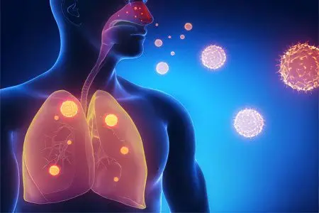
Infection. The pathogenetic process of pneumonia begins after the infection enters the lung tissue. Primary damage to the alveolar cells occurs, as a result, inhibition of the local protective function begins. This mechanism is called the heat stage, which is observed within approximately 72 hours from the moment of infection. During this time, the bacteria spread rapidly in the lungs, covering an increasing number of alveoli.
Burn. Damaged alveolocytes are impregnated with fibrinogenic fluid. The period of initial red hepatization is the transport of erythrocytes into the cavity of the alveoli. The lung is stained in a specific liver color. After completion of this process, the destruction of erythrocytes begins, attracting leukocytes to the site of injury. There comes a stage of gray hepatization. The color of the lung changes to grey. The entire hepatization process takes approximately 7-9 days.
Resolution. With adequate treatment, the disease ends in the resolution stage. Reconvalescence consists in cleansing the alveoli, restoring the physiological function of the lungs. The duration of the resolution period is affected by the severity of the clinical picture of pneumonia, possible complications.
The development of the pathogenesis of lung cancer corresponds to certain stages:
Initiation. At the beginning, the penetration of the pathogenic factor into the lung occurs. This stage of the development of the disease is reversible, since the destructive effect of the carcinogen on the DNA of the cell is just beginning, which leads to atypical transformations. If an oncogenic effect on the lung tissue is prevented during this period, then the further development of the disease can be completely stopped.
Promotion. Unfortunately, the first stage often goes unnoticed, so it is replaced by the promotion stage. Prolonged carcinogenic effects lead to more and more significant damage to the DNA, and the activation of genes responsible for the development of cancer occurs. Lung cells are transformed into atypical ones, acquire a differentiated structure and become capable of rapid reproduction.
Progression. The stage of progression usually corresponds to stage IV of the development of a cancerous tumor. At this stage, the formed neoplasm progresses, grows uncontrollably, involves not only nearby organs in the process, but also gives regional metastatic foci.
Tuberculosis pathogenesis:
The onset of tuberculosis corresponds to the moment when infected dust particles are inhaled by a person who is not immunized. Mycobacteria enter the lungs, where they are taken up by alveolar macrophages. If a person has specific immunity, then the disease does not develop.
Otherwise, Koch’s wand kills macrophages, the proliferation stage begins. The pathogen attacks immune blood cells, affects an increasing number of alveoli in the focus of initial introduction. Over time, mycobacteria move to the lymph nodes, systemic organs. After that, it is possible to form a powerful enough immune response to stop the disease.
The stage of formation of the immune response is characterized by the death of pathogenic bacteria. Most patients experience some residual effects but recover completely over time. With an unfavorable course of this period, tuberculosis progresses with the further formation of necrotic foci in the lung tissue – caverns. At this stage, the patient actively excretes the causative agent of tuberculosis into the environment along with saliva and sputum.
Signs by which pneumonia can be distinguished from lung cancer and tuberculosis
In order to correctly assess changes in the body, it is important to know the main differences between clinical manifestations.
Symptoms | Pneumonia | Lung cancer | Tuberculosis |
Disease onset | Sharp | Slow, a lot of time passes from the onset of the action of a carcinogen to a clear picture of the disease | Gradual, with the addition of more and more symptoms |
Temperature | It rises to very high values - above 39 ° C | Jumps up to 38 ° C, accompanied by bouts of weakness | Constantly elevated to 37-38°C |
Cough | Strong, at the beginning of the disease – dry. Over time, copious sputum appears | Appears during the process. As the disease progresses, it becomes paroxysmal, painful. May be permanent | Strong, with colorless sputum. The discharge is odorless. During coughing, small bubbling rales are expressed |
Blood in sputum | At the height of the disease, sputum acquires a rusty color due to the presence of red blood cells. | Appears at 2-3 stages of the oncological process | Blood impurities in the sputum appear at later stages |
Pain | Painful sensations in the chest appear when the pleura is involved in the inflammatory process, aggravated by coughing | absent at the onset of the disease. Appears over the course of the process, is not stopped by painkillers | Pain from the diseased lung increases during coughing |
Sweating | Not typical | No | Elevated, more pronounced at night |
Puffiness | No | Swelling of the tissues of the face, neck | No |
Diagnosis of pulmonary diseases
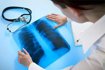
Among the diagnostic measures, hardware methods should be singled out. If earlier in the doctor’s arsenal there was only X-ray, now there is magnetic resonance, computed tomography. Scanning technologies make it possible to perform accurate images of the lungs, which show both diffuse lesions and emerging malignant processes.
Of the laboratory methods, a study of blood and sputum is mandatory. In addition to increasing the ESR and lymphocytes, they help to establish deviations of other elements of the blood formula. In addition, the biochemical composition of the blood, the level of albumins, globulin proteins are examined. Sputum culture allows you to confirm or exclude the presence of atypical cells, to identify specific pathogens.
Features of treatment
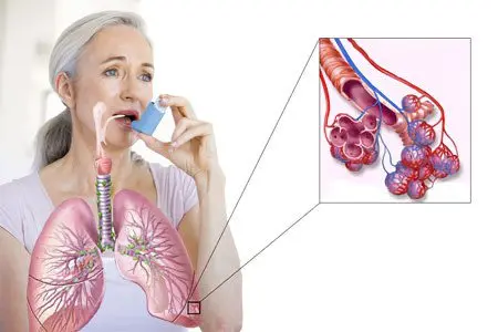
Regarding pneumonia, only medications are used:
Antibacterial.
Anti-inflammatory.
Expectorants.
All drugs are aimed at eliminating the causative agent of the disease and alleviating, suppressing negative processes in the lungs, and relieving symptoms.
In the treatment of lung cancer, like any oncological disease, the following treatment methods are used:
Surgical.
Chemotherapeutic.
Ray.
If the patient’s condition and the course of the pathology allow, then all of the listed methods are used. Complex treatment gives a high effect, allows you to stop the disease or significantly extend the life of the patient.
Treatment of tuberculosis is aimed at eliminating the pathogen – mycobacteria (Koch’s sticks). For this purpose, anti-tuberculosis chemotherapeutic drugs are prescribed. In the case of reversible forms of the disease, they can be used to completely cure the patient. If tuberculosis has passed into a severe, neglected form, the task of therapy is to stabilize the patient’s condition, to stop the release of bacteria along with biological fluids.
Pneumonia is a serious lung disease that requires hospitalization of the patient in a specialized hospital, which has the equipment necessary for artificial ventilation of the lungs. A correct and timely diagnosis significantly increases the patient’s chances for a quick full recovery.


