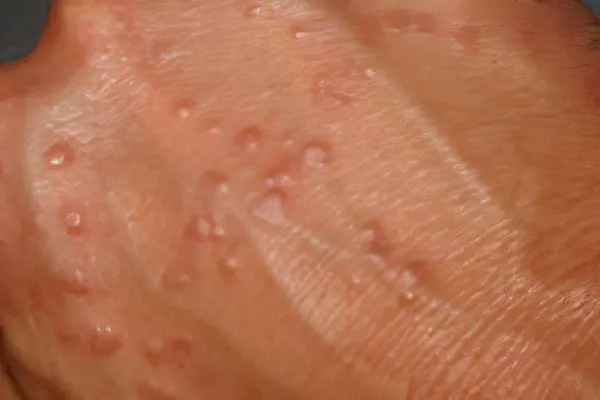
skin granuloma

The basis for the onset of skin granuloma may be a violation in the immune system of several types, such as hypersensitivity, allergic and cytological reactions. Granulomatous inflammation is an indicator of deviations in the protection of the immune system. Granuloma is classified, taking into account the reaction of tissues and the predominance of cells of various types in the inflammatory process, the presence of suppuration, necrotic type changes, the presence of foreign bodies, the presence of infectious agents.
– Tuberculoid (epithelioid-cellular) type of granuloma is observed in chronic infections. Diseases such as tuberculosis, actinomycosis, syphilis (secondary), rhinoscleroma and many others are the basis for the development of this type of granuloma.
– Granuloma of the sarcoid (histiocytic) type has, in its particularity, a reaction of tissues with a characteristic predominance of giant cells with many nuclei. It can develop in the presence of sarcoidosis, in the presence of tattoos on the body, with the introduction of zirconium.
– Necrobiotic (palisade-shaped) type of granuloma is characteristic in the presence of annular granuloma, lipoid necrobiosis, rheumatic nodules, venereal lymphogranuloma. The genesis of this type of granuloma can be different, including accompanied by a deep change in blood vessels. There is a type of granuloma, which is the result of a reaction of the skin to a foreign body, around which macrophages and giant cells of foreign bodies accumulate.
– Mixed type – combines signs of several types of granulomas.
Given the variety of types of skin granulomas, a dermatologist is able to make an accurate diagnosis after a complete examination, including a histological examination of skin samples, then an individual treatment is prescribed.
Granular faces
Facial granuloma, known as eosinophilic granuloma, is essentially a rare type of chronic dermatosis. Its manifestations can be single or multiple papules, as well as plaques that have a brown color. This disease is often observed by dermatologists, but its clinical picture is very diverse and diagnosis can be difficult. Granuloma may resemble other dermatoses – skin lymphoma, several types of lupus, sarcoidosis. Histology of a skin sample is the only method that allows an accurate diagnosis.
The cause of facial granuloma is not fully understood. Some authors tend to consider granuloma a late phase of leukoclastic type vasculitis, since they have a similar histology pattern. Facial granuloma is a chronic disease with a possible periodic exacerbation or remission. The illness can last from a couple of months to several years. Most often, the diagnosis of facial granuloma is made to patients after 45 years of age and most of them are men.
Internal organs in this disease, as a rule, do not suffer. Treatment of facial granuloma is aimed at cosmetic elimination of skin defects. In therapy, methods such as the antileprosy drug Dapsone, corticosteroid preparations in the form of ointments and injections, as well as PUVA therapy are used – treatment using a photoactive substance in conjunction with irradiation of the skin with long waves of ultraviolet radiation. Sometimes laser therapy, cryotherapy, dermabrasion are used. Only a doctor can choose a method.
Lip granuloma

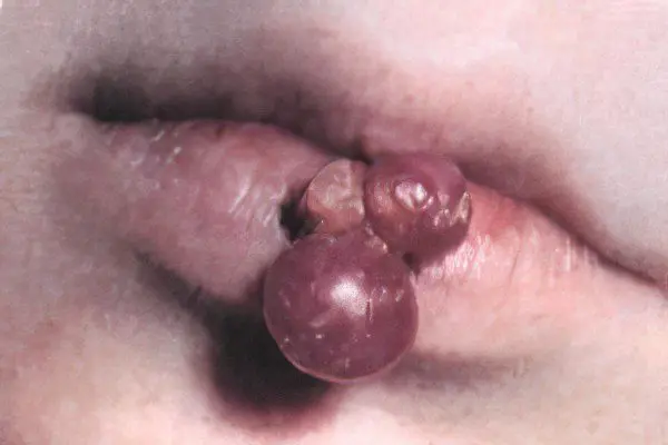
Lip granuloma (pyogenic granuloma) is a rapidly progressive disease that doctors refer to as a granulation hemangioma. This disease also affects the skin, mucous membranes, but a granular rash is common on the lips. It is not a tumor. The cause of the disease may be the body’s reaction to a mechanical injury, a hormonal disorder in the body, a consequence of treatment with retinoids (substances that are analogous to vitamin A and used in creams for problem skin), and other factors of influence are possible.
Typically, granuloma is observed in children, adolescents and adults aged not exceeding the age of thirty. Sometimes there is a development of the disease against the background of a violation of the vessels of the skin, which is manifested by the expansion of capillaries and looks like a wine stain. Also, a telangiectatic angioma can serve as a background for a granuloma, which manifests itself in newborns in the occipital region, forehead, neck, bridge of the nose, has a red-pink color and resolves on its own by the age of two.
Externally, a pyogenic granuloma may look like a red or purple dome-shaped papule, the surface of which is shiny and bleeds easily. Sometimes the papule may have a leg. The disease stretches for several months, and the focus increases as much as possible (over 15 mm) in 21 days, then it may decrease. Many experts in the presence of such a granuloma recommend a surgical method, since more gentle superficial removals provoke relapses due to incomplete removal of the tumor.


