Contents
What is a radius fracture?
Fracture of the radius – this is one of the most common household injuries, about 16% of all recorded acute pathologies of the skeletal system are precisely such injuries. Mankind has faced this type of fractures throughout its history, in burials more than 5 thousand years old, archaeologists find bones with traces of such injuries, and the first ancient, Egyptian, Chinese treatises known to us already contain recommendations for the treatment of such victims. This pathology is so widespread, due to the mechanism of its occurrence, the victim receives an injury by falling on an outstretched hand, or by a strong blow with an outstretched hand on something hard enough.
More often this injury occurs in women, after menopause, more than half of these injuries are received by them. This is due to the fact that during this period their calcium content in the bones decreases, and they become more fragile, and even a small load can lead to injury. Next, we will take a closer look at how such damage occurs, what symptoms it has, how to treat it, and how dangerous a fracture of the radius can be.
Fracture of the radius with displacement

A displaced fracture of the radius develops if the parts of the broken bone move relative to each other. The types of such fractures are very different, and differ in the direction and type of movement of damaged bone fragments, their localization, and the integrity of the skin.
There are several groups of such fractures:
Closed – all fragments of a broken bone are under the skin, they are the most favorable for the patient, the area of injury is sterile, the risk of possible complications is minimal, among fractures of this type.
Open – in which fragments of a broken bone rupture the skin, and the area of injury is in contact with the external environment, such a wound is not sterile due to microorganisms entering it from the external environment, such injuries are dangerous with possible infectious complications.
Intra-articular – the fracture line is completely or partially in the joint cavity, as a result, blood from the broken bone enters it, hemarthrosis develops, there is a significant risk of disrupting the normal operation of the damaged joint.
A change in the ratio of bones in the area of injury may be a consequence of the injury itself, for example, when the bone is crushed into fragments, or it may be the result of muscle work. This happens when they pull one end of the bone in their direction, and it will mix with another part of the bone to which this muscle is no longer attached. As a rule, with displaced fractures, both variants of the pathological process are observed simultaneously, which makes it difficult to ensure adequate restoration of limb function.
A characteristic external sign of a fracture with mixing is a change in the shape of the limb externally visible to the eye, a characteristic deformation is observed, however, it must be understood that changes externally visible to the eye in such an injury occur only with severe destruction of bone tissue, and are relatively rare.
Widespread is the transverse and longitudinal displacement of bone fragments. With this type of injury, a transverse or oblique fracture first occurs, which divides the radius into 2 parts. As a result, one of the parts of the bone under the action of the contracted muscles goes to the side, in this case, a transverse fracture with displacement is observed. If the fracture was longitudinal, then part of the bone fragments, under the influence of a traumatic effect, moves up the arm, and they seem to slide relative to each other. In most cases, the victims have both transverse and longitudinal displacement of bone fragments.
Less common is a displaced fracture called an impacted fracture. It looks like this, the patient falls on his arm, and one part of the radius seems to be hammered into another, the bone in this case is a bit like a telescopic antenna, in which one part of the bone enters the other.
Since the middle of the 20th century, among the fractures of the radius, the proportion of compression fractures has been growing. This is directly related to the spread of road transport and industrial equipment, and as a result, to an increase in the number of victims in accidents related to machinery. The mechanism of injury in such situations differs from that typical for this pathology, bone damage does not occur as a result of a fall or a blow with a hand, but as a result of the infringement of the limb between two metal surfaces, as a result of which the bone is crushed, as if it were in a vise. Such injuries are characterized by extensive soft tissue damage, and many small bone fragments at the site of injury.
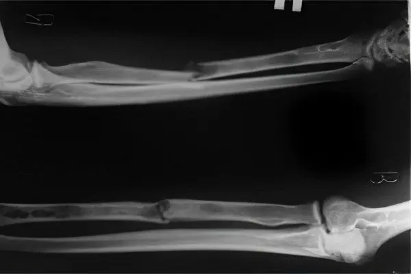
The main method of diagnosis of this type of fractures in modern medicine is an X-ray examination. A radiograph made in two projections allows the doctor to assess the position of the bones relative to each other, and the severity of the injury.
Fracture of the radius without displacement

At least half of the cases of fractures of the radius occur without displacement, since the muscle mass of the forearm is much smaller than on the lower limb, or on the shoulder, then with incomplete fractures, muscle strength is not enough to displace bone fragments relative to each other. In some cases, even a complete transverse fracture of the radius is not accompanied by displacement of bone fragments.
The most common variant of a fracture of the radius without displacement is a crack in the bone tissue. A crack in traumatology is usually called an incomplete fracture, when there are damages only to some part of the bone, but they do not extend to its entire thickness. As a rule, cracks are the result of household and sports injuries in relatively young people. Their bones are elastic and strong enough to withstand severe stress, and a complete fracture due to falls from a small height or blows is quite rare.
Externally, such a fracture manifests itself in the form of swelling and pain at the site of injury, unlike a fracture with displacement and an open fracture of the radius, there will only be swelling and possibly a hematoma at the site of injury. On the radiograph with this type of pathology, a full-fledged fracture line may not be observed, but only damage to the periosteum, and compaction of the bone tissue at the site of injury.
Fracture of the radius in a typical location
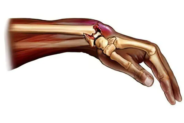
A fracture of the beam in a typical place is the most common injury to the radius, the destruction of bone tissue in this area occurs due to the anatomical features of the structure. In the area of the wrist joint, 3-4 cm from its articular surface, when falling on the hand, the maximum load occurs, and as a result, the bone does not withstand and collapses.
There are two main types of radius fracture in a typical location:
Colles fracture – is a hyperextension of the wrist joint, in which a fracture of the radius occurs in a typical place. In this type of injury, the distal (further down the limb) bone fragment blends towards the dorsum of the forearm. Approximately two-thirds of radius fractures in a typical location are of this type. For the first time, such a variant of the fracture was described in 1814 by Abraham Colles, a famous surgeon and anatomist who lived in Ireland.
Smith’s fracture – represents a flexion fracture of the radius, the victim in this case falls on the arm, the hand of which is bent towards the dorsum of the forearm. Thus, the distal bone fragment moves to the outer surface of the forearm. This type of typical injury to the radius was first described by Robert Smith in 1847. In fact, a beam fracture in a typical location is two types of fracture that mirror each other.
At present, a significant proportion of patients with a fracture of the beam in a typical location are women over 45 years of age. This is due to the consequences of menopause, which negatively affects the strength of bone tissue, and as a result, the resistance of bones to shock loads. An impact that at the age of 20 would only lead to a bruise, for a woman of 50 years old can easily end in a fracture.
The peak of appeals with such injuries in countries with a cold climate occurs in spring and autumn, this is due to ice, and an increase in the risk of falling, the number of people receiving bruises increases, and the number of fractures also increases.
Complications after a fracture of the radius

Complications of fractures of the radius can be divided into two large groups:
Immediate complications of injury are complications arising from the influence of injuries resulting from a bone fracture on the normal functioning of the limb.
Long-term consequences of an injury are complications resulting from incorrect treatment, or a violation of normal healing after an injury.
Immediate complications include:
Tears and injuries of the nerves that provide sensitivity or mobility of the limb. Bone fragments can, with their sharp edges, damage or tear large nerve trunks, depriving the area below the site of injury of signals from the brain. As a result, the ability to arbitrarily move the affected area may partially or completely disappear, sensitivity is lost.
Injuries to the flexor tendons of the fingers, bone fragments shifting towards the back surface of the forearm can damage the tendon bundle leading to the hand, and as a result, the victim completely or partially loses the ability to move the fingers of the hand.
Tight edema of Turner’s hand, as a result of which reflex immobility of the fingers develops, the patient cannot make arbitrary movements with them, but if he tries to move them, he experiences severe pain. Severe osteoporosis develops to the bones of the wrist and cyst.
Injury to large main vessels, followed by intracavitary hemorrhage, such damage can lead to the development of long-term complications.
Complete or partial rupture of muscles, or separation of muscles from the places of attachment to the bone tissue, leads to the impossibility of subsequent voluntary movements of that part of the limb, the movement of which was carried out by the affected muscle.
Acute infectious complications, with open fractures, an infection can get into the wound, which in turn can lead to the formation of acute osteomyelitis. This pathological condition manifests itself in the form of purulent fusion of bone tissue with high temperature and intoxication.
Long-term effects of trauma include:
Ischemic contracture is a violation of the mobility of the joints of the affected limb due to an incorrectly applied plaster cast, which compresses the soft tissues, disrupting the blood supply, and as a result, adhesions are formed that impair the mobility of the joints involved.
Violations of the bone structure due to inadequate reposition, an incorrectly applied plaster cast, may not hold the bone fragments well enough, and during the time required for healing, they will take the wrong position, and in this position they will be fixed by the growing bone tissue.
Long-term infectious complications, as a rule, manifest themselves in the form of the formation of chronic osteomyelitis. This chronic purulent-septic disease develops due to the penetration of an infectious agent into the bone tissue, which, in the course of its vital activity, begins to gradually destroy the bone tissue, forming purulent cavities in the bone. The presence of these cavities causes intoxication, pain in the affected bone, and can lead to a pathological fracture, due to a decrease in the strength of the bone tissue in the affected area.
Long-term consequences of hemarthrosis, in the presence of an articular fracture of the radius inside, blood inevitably enters the joint cavity. The blood in the joint leads to the formation of a fibrin clot, and this protein aggregation links the joint surfaces together from the inside, and the person can no longer freely, fully bend the affected joint.
Edema after a fracture of the radius
Edema at the site of injury is a typical sign of a bone fracture, and injury to the radius is no exception. Let’s take a closer look at how dangerous it can be with such a fracture, and what to do with it. In most cases, swelling does not pose a significant danger, but it should not be taken lightly.
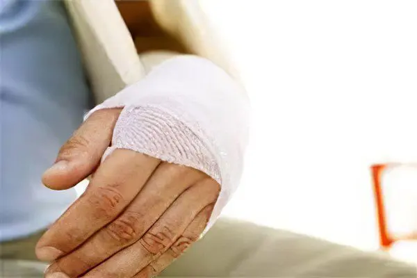
If you do not take into account the magnitude of the growing edema when applying a plaster cast, then its increase in the closed space of the plaster splint will lead to tissue compression and ischemia, which, in turn, can cause the formation of ischemic contracture.
An equally dangerous complication is Turner’s tight edema, as a result of which the patient loses the ability to move the hand, and without timely medical attention this can lead to a long-term loss of mobility in the affected joints.
You should carefully monitor the condition of the hand and tissues visible from under the plaster splints, since the presence of edema under the bandage is difficult to identify, and its long existence is dangerous not only with ischemic, but also with thromboembolic complications. That is, in the area of edema, due to a slowdown in blood flow, blood clots can form, which can later move through the vessels and lead to serious health problems.


