


Contents
Forearm
The forearm is a part of the upper limb located between the elbow and the wrist.
Anatomy of the forearm
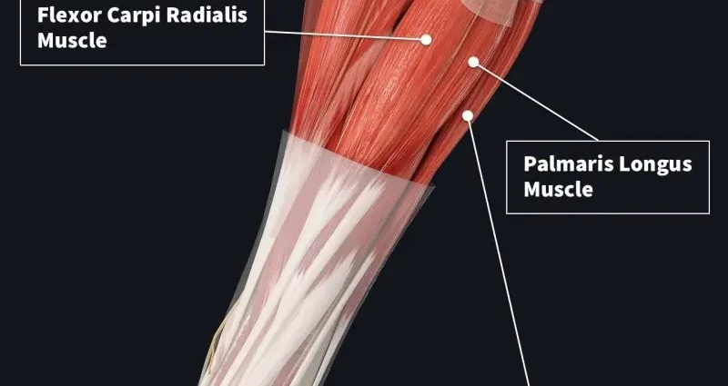
Structure. The forearm is made up of two bones: the radius and the ulna (commonly known as the ulna). They are linked together by an interosseous membrane (1). About twenty muscles are arranged around this axis and are distributed through three distinct parts:
- the anterior compartment, which brings together the flexor and pronator muscles,
- the posterior compartment, which brings together the extensor muscles,
- the external compartment, between the two preceding compartments, which brings together the extensor and supinator muscles.
Innervation and vascularization. The innervation of the forearm is supported by three main nerves: the median and ulnar nerves at the anterior compartment and the radial nerve at the posterior and lateral compartments. The blood supply to the forearm is mainly carried out by the ulnar artery and the radial artery.
Forearm movements
The radius and ulna allow forearm pronosupination movements. 2 Pronosupination is made up of two distinct movements:
- The supination movement: orient the palm of the hand upwards
- The pronation movement: orient the palm of the hand downwards
Wrist and finger movements. The muscles and tendons in the forearm extend to form part of the musculature of the hand and wrist. These extensions give the forearm the following movements:
- abduction and adduction of the wrist, which therefore respectively allow the wrist to move away from or approach the body
- flexion and extension movements of the fingers.
Pathologies of the forearm
fractures. The forearm is often the site of fractures, whether of the radius, ulna, or both. (3) (4) We find in particular the Pouteau-Colles fracture at the level of the radius, and that of the olecranon, part forming the point of the elbow, at the level of the ulna.
osteoporosis. Loss of bone density and increased risk of fractures in people over 60 years old.
Tendinopathies. They designate all the pathologies that can occur in the tendons. The symptoms of these pathologies are mainly pain in the tendon during exertion. The causes of these pathologies can be varied. In the forearm, epicondylitis, also called epicondylalgia, refers to pain appearing in the epicondyle, a region of the elbow. (6)
Tendinitis. They refer to tendinopathies associated with inflammation of the tendons.
Forearm treatments
Medical treatment. Depending on the disease, different treatments may be prescribed to regulate or strengthen bone tissue or reduce pain and inflammation.
Surgical treatment. Depending on the type of fracture, a surgical operation can be performed with, for example, the placement of pins, a screwed plate or even an external fixator.
Forearm examinations
Physical examination. Diagnosis begins with an assessment of forearm pain to identify its causes.
Medical imaging examination. X-ray, CT, MRI, scintigraphy or bone densitometry examinations can be used to confirm or deepen the diagnosis.
History and symbolism of the forearm
External epicondylitis, or epicondylalgia, of the elbow is also referred to as “tennis elbow” or “tennis player’s elbow” since they occur regularly in tennis players. (7) They are much less common today thanks to the much lighter weight of current rackets. Less frequent, internal epicondylitis, or epicondylalgia, are attributed to the “golfer’s elbow”.