Contents
In line with its mission, the Editorial Board of MedTvoiLokony makes every effort to provide reliable medical content supported by the latest scientific knowledge. The additional flag “Checked Content” indicates that the article has been reviewed by or written directly by a physician. This two-step verification: a medical journalist and a doctor allows us to provide the highest quality content in line with current medical knowledge.
Our commitment in this area has been appreciated, among others, by by the Association of Journalists for Health, which awarded the Editorial Board of MedTvoiLokony with the honorary title of the Great Educator.
Quite often, young mothers visit a doctor because they are afraid to touch the baby’s head while bathing, because: “there are some soft holes on it that can damage the brain”. These “holes” are the fontanel. There are several of them, but the most important ones are the front cap (large) and the posterior cap (small).
The bones of a newborn’s skull are not fully fused together, as in an adult. This is because the baby’s head continues to grow after birth. The flexible connections of the skull bones also enable the passage of the fetal head through the birth canal during natural delivery. The bones of the skull then overlap each other in a tiled shape, reducing the circumference of the head. Up to a few days after giving birth, individual bones return to their place; between them there are gaps of several millimeters, called seams.
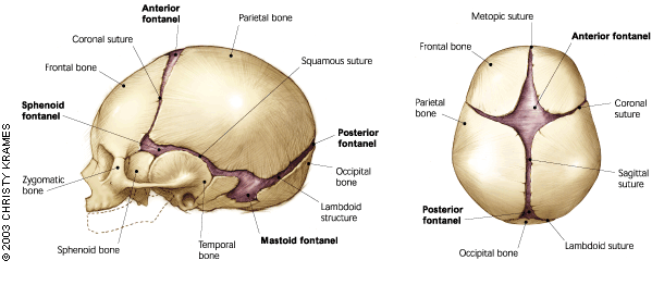
Fontanelle – structure of an infant’s head
Immediately after delivery, a newborn’s head is usually slightly distorted, often elongated or asymmetrical. It’s the effect of squeezing through the narrow birth canal. The longer a baby stays upside down, the greater the likelihood of an asymmetrical skull shape after delivery. Therefore, children who are born via caesarean section much less frequently have distorted or elongated heads.
Of course, the asymmetry in the newborn wears over time and the shape of the head takes on standard dimensions.
The average circumference of a newborn’s skull is approximately 34 centimeters. The baby’s cranial sutures are very loosely connected with each other, thanks to which the head can freely grow. Importantly, the bones of a child’s skull grow together all the time until they reach adulthood. However, they do not fuse simultaneously. Certain norms of values have been set, which can be achieved by the circumference of a newborn’s head. Norms and percentile grids are used to measure the child during visits to the pediatrician. These measurements are especially important in the first year of a baby’s life. The pediatrician then checks whether the head grows at the correct pace and whether there are any abnormalities in its growth.
You can find more information about caring for a newborn baby here: Newborn – concerns, breastfeeding, care, care
Fontanelle – characteristics and basic information
The fontanel is the non-ossified symphysis that connects the skull bones of newborns and infants. The fontanels are filled with connective tissue membranes. The membranous connection between the bones of the skull makes it easier for the baby to get out into the world. The fontanelles ensure greater flexibility of the skull and thus enable the child to position properly before birth. In addition, thanks to the membranous structure of the fontanelle, it is possible to perform ultrasound examinations and assess the condition of the child’s brain. This examination is indicated in the diagnosis of intracerebral bleeding or in the detection of congenital defects and other abnormalities of the skull.
What is worth knowing about the structure of the skull? Check it out: Skull bones – everything you need to know
Types of fontanels
There are several fontanels in newborns, although the anterior and posterior fontanels are considered the most important. The following types of fontanelles are listed:
- front fontanel – it has the shape of a diamond or a kite. It is located in the midline of the skull, i.e. at the intersection of the coronal suture, the sagittal suture and the frontal suture between the frontal bones and the parietal bones. It can be of various sizes, with preterm babies being the largest. The fontanel dimensions range from approximately 2 × 2 cm to 3 × 5 cm. It is soft and flat and can be felt very well. In a few months old baby, especially when crying, you can feel its pulsation, which is the norm;
- posterior fontanel – the posterior crown is located in a straight line from the large parietal, behind the top of the head at the junction of the sagittal suture and the parietal suture, between the occipital bone and parietal bones. It has the shape of a triangle about 1 cm wide. It happens that at the time of birth it is already overgrown;
- lateral fontanel – are also called sphenoid and nipple fontanels. It is a fontanel that occurs ungrown in premature babies. These fontanelles become overgrown in utero. The sphenoid, or anterolateral, fontanel is located between the sphenoid, parietal, temporal and frontal bones. On the other hand, the mammary gland, or posterior-lateral, is located between the temporal, parietal and occipital bones.
If you want to learn about the baby’s development in the mother’s womb, check out: Fetal development stages – first, second and third trimesters
The role of fontanel in an infant’s skull
The fontanel plays a very important role in the baby’s head. Among other things, two main functions can be distinguished:
- facilitating childbirth – the fontanelles make it easier for the baby to come into the world. The circumference of the fetal head is quite large, which makes it difficult for a baby to squeeze through the birth canal. Due to the fact that the bones of the skull are not fused, they do not break during childbirth, but move slightly overlapping each other;
- facilitate research – neonatologists who test infants define fontanel as windows or eyes in the head that allow them to examine the normal functioning of the nervous system. Thanks to the fontanelles, it is possible to perform trans-epithelial ultrasound.
Read more about how to spot your upcoming birth: Signs of labor
Fontanelle – the most common problems
The health of the infant can be inferred from the appearance of the front crown. The problems that parents report to pediatricians usually concern:
- collapsed fontanell – may be a sign of dehydration. The collapsed fontanel usually occurs in such conditions as: chronic diarrhea, severe vomiting or fever. In addition to the physical examination, laboratory tests are also performed, i.e. blood count, ion concentration or general urine test;
- convex fontanel – may be a symptom of meningitis. The fontanel in this case is convex and pulsating. Changes in the fontanel’s appearance are accompanied by fever and severe general condition of the child. The bulge of the fontanel, without fever and pulsation, indicates an increase in pressure inside the skull, which may be a symptom of hydrocephalus or subdural hematoma. Usually, the cranial sutures also widen;
- pulsating fontanel – this symptom should not be alarming for parents. The pulsation of the fontanel usually occurs when the baby is crying. As the crying stops, the throbbing stops. A pulsating fontanel is considered normal;
- pulsating fontanel – if the child also has seizures, drowsiness or apathy, this may mean that the child is developing an infection.
More information about diseases that most often affect newborns can be found here: The most common diseases of infants – colic, constipation, ear infections
When does the fontanel flare up?
Growing up of fontanels is a dynamic process. The front fontanel grows between 9 and 18 months of age, and the rear – already between 6 and 8 weeks of age. However, the variability of the fontanel overgrowth time between infants is considerable.
It happens, however, that the front fontanel grows too early. If it is accompanied by overgrowth of cranial sutures, as evidenced by rolled thickenings along their course, the so-called craniostenosis – a disease involving the inability to enlarge the circumference of the head. Surgical treatment is then necessary, as too small a skull prevents the growth and proper development of the brain.
Fontanelle – examination
The fontanel should always be under the supervision of a pediatrician. During follow-up visits to the pediatric office, the doctor should measure the circumference of the child’s head and examine the fontanel. The pediatrician measures the size of the fontanelles and assesses whether they are concave, convex or pulsating. The size of the fontanelle affects the recommended dose of vitamin D. Too early fontanel growth may be the result of too much vitamin D supplementation. Therefore, the dosage of vitamin D depends on the age, weight and development of the child. In addition, the size of the fontanelle helps in determining the infant’s age. The membranous structure of the fontanell, in turn, allows the doctor to perform an ultrasound examination and assess the condition of the child’s brain.
Attention! An abnormal fontanel size may indicate diseases such as: diseases of the skeletal system, genetic syndromes, endocrine or metabolic disorders, and congenital pathologies of the central nervous system. On the other hand, the fontanel which grows too late may indicate rickets or malnutrition of the child.
How to care for the fontanel?
Often, parents fear that touching the fontanelles may harm their child. This is due to the apparent delicacy of this structure. It turns out, however, that the fontanel and the tissue that builds it are strong, so you shouldn’t avoid combing your baby’s hair, or even washing the head. Caring for the baby’s head should be the same as caring for the rest of the newborn’s body. However, remember to observe the fontanel during care, as it will allow for early detection of irregularities. Similarly, if erythema-exfoliating lesions appear on the child’s head, use an olive that softens the skin or consult a pediatrician on the selection of appropriate cosmetics for the care of the baby.


