Contents
In line with its mission, the Editorial Board of MedTvoiLokony makes every effort to provide reliable medical content supported by the latest scientific knowledge. The additional flag “Checked Content” indicates that the article has been reviewed by or written directly by a physician. This two-step verification: a medical journalist and a doctor allows us to provide the highest quality content in line with current medical knowledge.
Our commitment in this area has been appreciated, among others, by by the Association of Journalists for Health, which awarded the Editorial Board of MedTvoiLokony with the honorary title of the Great Educator.
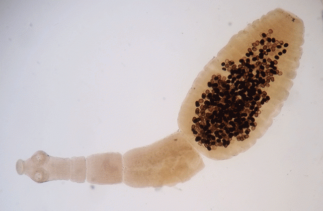
Echinococcosis is a parasitic disease that occurs in humans in two main forms: single-chamber echinococcosis (also known as echinococcosis) and multi-chamber echinococcosis (alveococcosis), caused by tapeworms Echinococcus granulosus and Echinococcus multilocularis, respectively. The infection is spread through the consumption of food or water containing the parasite’s eggs, or through close contact with an infected animal. The disease often begins asymptomatically and may last for years. Echinococcosis usually starts in the liver but can spread to other parts of the body, such as the lungs or the brain. The disease occurs in most parts of the world and currently affects around one million people.
- Echinococcosis is a parasitic disease. It develops in secret at first – symptoms often only appear after many years
- The infection most often occurs after consuming contaminated water or fruit, e.g. forest berries
- Malaise, abdominal pain, nausea are symptoms of echinococcosis in humans
- You can find more such stories on the TvoiLokony home page
Echinococcosis – causes
The causes of single-chamber echinococcosis
People who accidentally swallow tapeworm eggs Echinococcus granulosusare at risk of infection. Dogs that eat sheep slaughtered at home and other livestock contract the tapeworm Echinococcus granulosusand tapeworm eggs can be found in their stools. Direct contact with infected dogs, especially between children and their dogs, can lead to human infection. The consumption of soil, water, and vegetables contaminated with dog’s faeces can also lead to infection. It is worth adding that eggs Echinococcus granulosus they can survive in snow and frost.
People can become infected with parasite eggs by transferring them “from hand to mouth”.
- By ingesting food, water, or soil contaminated with feces from infected dogs. This may include grass, herbs, vegetables, or berries harvested from the fields.
- By stroking or touching dogs infected with a tapeworm Echinococcus granulosus. Dogs can shed tapeworm eggs in their stools and their fur can become contaminated with them.
You can buy a mail-order study at medonetmarket.pl – check the offer HERE
The causes of multi-chamber echinococcosis
People who accidentally swallow tapeworm eggs Echinococcus multilocularisare at risk of infection. In this case, high-risk individuals include trappers, hunters, vets, or others who come into contact with wild foxes, coyotes or their droppings, or dogs and domestic cats that have the ability to eat wild rodents infected with echinococcosis. People can become infected with parasite eggs in the same way as in the previous case.
- By directly eating food contaminated with fox or coyote faeces. This may include grass, herbs, vegetables, or berries harvested from the fields.
- By stroking or touching dogs or domestic cats infected with the tapeworm Echinococcus multilocularis. These animals can shed tapeworm eggs in the stool and their fur can be contaminated with them. Some dogs wallow in foreign material (such as wild animal faeces) and can become contaminated this way.
Also read: Veterinary diseases. What can a special task doctor suffer from?
Echinococcosis – symptoms
Symptoms of single-chamber echinococcosis
Human infection by single-chamber tapeworm (E. granule sus) leads to the development of one or more echinococcal cysts, located most often in the liver and lungs, and less frequently in the bones, kidneys, spleen, muscles and the central nervous system.
The asymptomatic brooding period can take years for echinococcyst cysts to develop clinically symptomatic, but about half of all patients who receive treatment for the infection do so within a few years of initial parasite infection. Abdominal pain, nausea, and vomiting are often seen when wheals appear in the liver. When lung involvement, clinical signs include chronic cough, chest pain, and shortness of breath. Other symptoms depend on the location of the echinococcal cysts and the pressure exerted on the surrounding tissues. Non-specific symptoms include anorexia, weight loss, and weakness.
Symptoms of multi-chamber echinococcosis
Multichamber echinococcosis is characterized by an asymptomatic incubation period of 5–15 years and a slow development of the primary tumor-like lesion that is usually found in the liver. Clinical symptoms include weight loss, abdominal pain, general malaise, and signs of liver failure. Metastasis can spread to organs adjacent to the liver (such as the spleen) or distant sites (such as the lungs or the brain) after the parasite has spread through the blood and lymphatic system. Unfortunately, untreated multichamber echinococcosis is progressive and fatal.
Echinococcosis – diagnosis
Computed tomography, MRI, and ultrasound of the abdominal cavity may be pathognomonic for single-chamber echinococcosis in the liver if cysts and alveolar sand are present, but simple cysts may be difficult to distinguish from benign cysts, abscesses, or malignant neoplasms. The presence of bubble sand in the aspirated cystic fluid is diagnostic. The World Health Organization (WHO) criteria use the imaging results to categorize cysts as active, transient, or inactive. Lung involvement may appear as round or irregular changes in the lungs on a chest X-ray. Multichamber echinococcosis usually presents itself as an invasive tumor.
Serological tests (enzyme immunoassay, indirect hemagglutination test) are sensitive in detecting infections, which can be confirmed by demonstrating echinococcosis antigens by immunodiffusion or immunoblot tests. A complete blood count can detect eosinophilia.
See also: Signs that death is just around the corner. 12 signals sent by the human body
Echinococcosis – treatment
Treatment of single-chamber echinococcosis varies depending on the type, location, and number of cysts, and whether the imaging results indicate that the cysts are active, transient, or inactive.
Surgical resection can be curative and is the best treatment for complicated lesions with the following characteristics:
- ruptured cysts;
- cysts with biliary fistulas;
- cysts oppressing vital structures;
- cysts with descendant cysts;
- cysts> 10 cm in diameter;
- superficial cysts with a risk of rupture due to trauma;
- cysts with accompanying extrahepatic disease.
In the case of small (
Small, single-chamber cysts can be treated with albendazole alone for several months, resulting in a 30% cure. Albendazole itself is also a treatment method for inoperable cysts.
Only observation is an option for asymptomatic cysts that are naturally inactivated (not inactivated by drug treatment). In the case of echinococcosis, there have been cases where a liver transplant has saved the lives of several patients.
The dose of albendazole is 400 mg orally 2 times a day (7,5 mg / kg 2 times a day in children up to a maximum of 400 mg 2 times a day). The second choice is mebendazole at a dose of 40 to 50 mg / kg body weight per day.
Patients with E. multilocularis should receive albendazole (in the above daily doses used for single-chamber echinococcosis) for ≥ 1 week, followed by surgical resection if possible, depending on the extent, location and symptoms of the lesion. The prognosis is poor unless all larval mass can be removed. Albendazole is given continuously for at least 2 years and patients are monitored for relapse for 10 years or more.
Unfortunately, prolonged therapy with high doses of albendazole can cause bone marrow suppression, liver toxicity, and temporary hair loss. It is important to monitor complete blood count and liver enzymes during use.
Also read: How to get rid of parasites from the body?
Echinococcosis – prophylaxis
There are several different steps you can take to prevent echinococcosis. In areas of the world where the parasite is common, education can help.
- Removing worms from dogs and cats can help contain the spread of infection;
- Proper disposal of animal faeces can reduce exposure to tapeworm eggs;
- It is also very important to thoroughly wash your hands after handling dogs, cats, or objects that may be contaminated with their faeces (e.g. garden soil). It is also not recommended to let dogs lick our face;
- Preventing dogs from catching rodents;
- Fencing of property (garden, vegetable garden) to prevent access of foxes and other wild predators;
- Constant supervision of children during their stay in the forest (the point is that they do not raise anything to their mouths or play with animals);
- Proper handling of cattle on farms and slaughterhouses is also essential. This includes the enforcement of meat inspection procedures;
- Avoiding undercooked or raw beef, pork, and fish can also help avoid echinococcosis;
- Also, washing fruit and vegetables, especially in areas where tapeworms are common (especially fruit and vegetables from plots where predators may have access), can help prevent infection.
- Thorough hand washing, especially after returning from mushroom picking or forest hikes;
- Not using water from tanks accessible to animals for washing fruit and vegetables or for consumption;
- Regular deworming of dogs and cats (this especially applies to farm animals raised in the countryside).
- Regular deworming of dogs, either permanently or temporarily in endemic areas, with preparations containing praziquantel is recommended.
Echinococcosis – occurrence
Single chamber echinococcosis occurs worldwide on every continent except Antarctica. In turn, multi-chamber echinococcosis is limited to the northern hemisphere, in particular to the regions of China, Our Country, and countries in continental Europe and North America.
The highest prevalence is in rural areas where older animals are slaughtered. Depending on the species infected, losses in livestock production attributed to echinococcal echinococcosis may include reduced carcass weight, reduced skin value, reduced milk production, and reduced fertility.
In Poland, echinococcosis was first detected in 1994 in the province. Pomeranian Voivodeship at foxes. Some time later, in 2007, 40 human cases were reported, and in 2008 about 20 cases were recorded in the following voivodships: Podkarpackie, Warmińsko-Mazurskie and Pomorskie.


