Contents
In line with its mission, the Editorial Board of MedTvoiLokony makes every effort to provide reliable medical content supported by the latest scientific knowledge. The additional flag “Checked Content” indicates that the article has been reviewed by or written directly by a physician. This two-step verification: a medical journalist and a doctor allows us to provide the highest quality content in line with current medical knowledge.
Our commitment in this area has been appreciated, among others, by by the Association of Journalists for Health, which awarded the Editorial Board of MedTvoiLokony with the honorary title of the Great Educator.
In a healthy person, both eyeballs move in different directions thanks to the work of the muscles that put each eye in motion. While looking, we reflexively position our eyes so that the image of the object falls symmetrically on the macular part of the retina simultaneously in both eyes. Disturbances in the position and movement of the eyes may appear as a result of neurological ailments that cause paralysis of the gaze.
Disorders of positioning and eye movements – definition
Normally our eyeballs move in different directions thanks to the work of the muscles that move each eye. On the other hand, disturbances in the position and movement of the eyes may result from improper centers responsible for the movement of the eyes due to e.g. neurological ailments (then the gaze is paralyzed). Muscle paralysis also occurs as a result of injuries and infections that require ophthalmic treatment.
Causes of eye movement and positioning disorders
Disturbances in normal eye movement may occur due to:
- diseases or injuries of the central nervous system:
– multiple sclerosis,
– aneurysm,
– tumor,
— myasthenia,
– infectious diseases,
– Lyme disease,
– poisoning.
In addition, the disease is due to:
- hemangiomas,
- arterial hypertension,
- diabetes
- muscle diseases,
- tumors,
- injuries,
- inflammatory changes,
- retrobulbar hematoma.
General symptoms of impaired position and eye movement
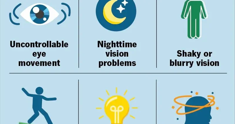
The symptoms of this ailment (regardless of the cause) include:
- black,
- double vision, which becomes worse when the eye moves towards the paresis muscle
- compensatory head position (appears when the muscle paresis is minor),
- limited or missing eye movement in the direction that the affected muscle is moving it,
- incorrect arrangement of elements in the surrounding space.
Strabismus in children – Already in the first year of life, it is a separate issue. The most common cause is refractive error, especially hyperopia and ataxia. Convergent strabismus usually occurs, caused by the excessive accommodation of the refractive eye. In the case of myopia, divergent strabismus develops. Sometimes the eyes cross alternately.
Types of eyeball paralysis
1. Paralysis of associated gaze – the eyeballs are unable to move in a given direction and are forced to turn in the opposite direction. Double vision does not occur in combination paralysis (as opposed to oculomotor paralysis). Persistence of paralysis for a long time leads to changes in other muscles of the affected eye and the other eye.
2. Paralysis of the oculomotor nerve – characterized by upper eyelid drooping, the eye is set in the divergent strabismus lower than the healthy eye. Movement of the eye up and down and towards the adduction is also impaired. The eye is unable to move downward (it only performs the abduction movement). Additionally, there is double vision and dilation of the pupils that do not respond to any light and do not constrict.
3. Block nerve palsy – is characterized by paralysis and paresis of the upper oblique muscle. The movement of the eyeball is significantly limited downward and in adduction. In addition, the movement of turning the eye inward is impaired. The diseased eye is slightly convergent and located higher than the healthy eye. There is also double vision, which intensifies especially when looking down, then double vision and spreads vertically. The patient’s head is tilted towards the healthy eye, as is the face, and the chin is lowered.
4. Palsy of the abduction nerve – causes the diseased eye to be in convergent strabismus. The patient develops a split image. The position of the patient’s head is characterized by turning the face towards the affected eye, where double vision is most intense.
Diagnosis of disorders of positioning and eye movements
Patient diagnostics is primarily focused on finding the cause of the disorders. For this purpose, a number of neurological, endocrine, internal and internal examinations as well as imaging of the brain and eye sockets are performed.
- Examination of the movement and position of the eyes – is a method that allows to determine which muscle has been affected. The doctor assesses how the eyeballs move in nine gaze directions (straight, right, left, oblique, up or down);
- The study of false localization in space – also allows to assess what paralysis of the eyeballs we are dealing with and which muscle is impaired;
- Double vision examination – is the most accurate method even in cases where the paralysis is minor and impossible to diagnose using other methods. This type of examination allows you to determine in which direction there is the greatest distance between the images. This is important because this direction is actually the direction of the defective muscle.
Treatment of disorders of positioning and eye movements
A quick start of treatment plays an important role in preserving the eyesight of the strabismus and therefore even the early symptoms of strabismus should not be underestimated, when the deviation is only periodic. Therapy is implemented based on the cause that caused the disorder. Treatment should be dealt with by a specialist in endocrinology, neurosurgeons, maxillofacial surgeon, ophthalmologist or ENT specialist.
Paralysis of the muscles of the eyeball can be very different, and regeneration also differs from patient to patient. At the beginning of the disease, doctors recommend exercising the oculomotor muscles to reduce the over-activity of the paralyzed muscle. After about two months of paralysis, botulinum toxin A is administered directly to the affected muscle. This is to reduce strabismus and get rid of double vision.
The treatment of ailments also involves the selection of appropriate eyeglass lenses that improve the vision defect and the use of various methods of covering the eyes and exercises to prevent the fixation of strabismus (especially permanent paresis). Sometimes it is necessary to surgically treat the ocular muscles. Treatment requires patience and discipline on the part of both the child and the caregivers – but it is essential for results.
Congenital eye movement disorders have a different course and have a different clinical picture. They occur, among others, from Moebius syndrome or Brown syndrome and unilateral paralysis of the muscles lifting the eyeball. Then there are also other symptoms that do not affect the eyes, while treatment is based on the cooperation of many specialists.


