Hair is considered a cultural ornament and its biological role is small. In addition, they are a sensory organ that senses a gentle touch, and also protects against solar radiation. For many people, hair is a very important element in shaping their own image, so any change in their number or structure may be a reason for visiting a doctor.
Hair diseases
Hair diseases often occur with changes in the area of the nail plates and skin diseases, and often indicate systemic disorders. The total number of hair follicles is about 5 million (about 100 on the head), and their density is 40-800 per cm2. The regulation of hair growth is influenced by hormonal factors (androgens) and genetic factors:
- racial features,
- color,
- family and gender-specific hair growth.
The anatomical structure of the hair is influenced by the root located in the hair follicle and the stem that comes out to the surface of the skin.
In order to maintain healthy and shiny hair, it is worth using proper care. At Medonet Market, you can buy a set of hemp cosmetics, the natural composition of which gently cares for the hair and sensitive scalp.
Developmental phases of hair follicles
- Phase I – growth (anagen);
- Phase II – physiological loss of life lasting up to two weeks (catagen);
- Phase III – the period of the dwell’s rest, which lasts 2-4 months (telogen).
Scalp diseases – types
1. Baldness
Alopecia can be the result of temporary or permanent hair loss in a limited area or all over the scalp (sometimes it can also affect other hairy areas). We can divide them into: non-scarring alopecia – physiological alopecia (infancy, puberty and menopause), androgenic alopecia (women and men), alopecia areata; scarring alopecia.
The reasons
The mechanism of alopecia may be related to either premature termination of the anagen phase (telogen effluvium) or damage to the anagen hair (anagenic or dystrophic alopecia), or both types of hair cycle disorders are observed.
The most common factors contributing to hair loss:
a. stress,
b. diet (deficiency of iron, folic acid, vitamins),
c. diseases with fever,
d. pregnancy (postpartum alopecia),
e. chronic systemic diseases (systemic lupus erythematosus, ulcerative colitis, diabetes, liver diseases),
f. drugs (cytotoxic and immunosuppressive agents, anticoagulants, beta-blockers, hormones, retinoids, anticonvulsants
and anti-thyroid, lipid-lowering, gold),
g. intoxication with heavy metals (thallium, mercury, arsenic),
h. acute infectious diseases,
i.mechanical (stretching, hair pulling),
j. surgery, blood loss,
k. hormonal disorders (hyperthyroidism, hypothyroidism).
2. PACKAGE HAIR LOSS (Fig. 20.1 and 20.2)
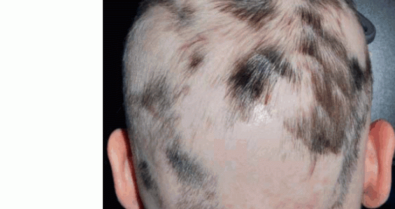
Alopecia areata is a focal, often progressive, hair loss without scarring. It affects both the scalp and other parts of the body. Single or multiple outbreaks appear suddenly, without obvious subjective symptoms (sometimes there is a slight inflammation, itching or burning skin).
Frequency of appearance
The prevalence of alopecia areata is estimated at 0,1-0,2% of the population, without any gender dominating, while approximately 30% of patients are children. It can present at any age, usually up to the age of 40, but the peak incidence is between 10-19. year
The causes of the disease
The etiopathogenesis of alopecia areata has not been fully elucidated. First of all, the role of autoimmune processes against one’s own hair follicles, which determine genetic predisposition, as well as not fully known environmental factors (e.g. stress), is emphasized. This is evidenced by the frequent coexistence of autoimmune diseases, such as Graves’ disease, Hashimoto’s goiter, vitiligo and diabetes.
Types and symptoms of alopecia areata
Alopecia areata can occur in the form of:
a.focal alopecia areata (alopecia focalis) – oval or round foci located on the scalp,
b. banded alopecia (ophiasis) – concerns the perimeter of the hairy skin in the occipital and temporal areas,
c. total baldness (alopecia totalis) – complete loss of scalp hair, eyebrows and eyelashes,
d. generalized alopecia (alopecia universalis) – loss of hair from the entire body,
e. malignant alopecia (alopecia maligna) – a form of generalized alopecia, characterized by a long course and resistance to treatment. In the course of AA, changes in the nail plates are often observed (punctured dimples, thinning and grooving).
FIG. 20.1. Alopecia areata of the eyebrows and eyelashes
FIG. 20.2. Alopecia areata
Diagnosis
During diagnostics, the presence of shallow miniature hairs is revealed (performed only in the event of diagnostic difficulties). The trichogram shows hair of reduced pigmentation, with a distinct constriction, taking the shape of an exclamation point (dystrophic and telogenous).
Alopecia areata should be differentiated from other ailments:
- toxic (drug-induced) alopecia,
- syphilitic alopecia,
- hair pulling (trichtillomania),
- mycosis of the scalp.
How to heal?
Local treatment: contact immunotherapy (topical application of strong contact allergens, ie DPCP-diphencyprone to induce an allergic reaction), cignolin (irritating), topical and intralesional steroids, 2-5% minoxidil solution (often in combination with cignolin).
General treatment: photochemotherapy (psoralens in combination with UVA irradiation) or UVB phototherapy, corticosteroids (only in cases of rapidly progressive, large areas), cyclosporine A, neurotrophic drugs, psychotherapy.
What’s the prognosis?
Most patients have a partial or complete spontaneous remission after about a year. In 10% of patients, the course of alopecia areata is chronic, extensive (the number of alopecia foci increases and hair loss affects all body surfaces) and is resistant to treatment.
3. ANDROGENIC ALOLINE
Androgenetic alopecia (AGA) is a progressive, androgen-dependent hair loss process that follows a closely programmed genetically programmed pattern. This ailment is the most common cause of hair loss in both women and men – it covers about 95% of all cases of the disease.
The causes of the disease
The causes of androgenetic alopecia include:
- genetic predisposition (autosomal dominant inheritance with varying degrees of gene penetration),
- age (onset between 12 and 40 years of age, and in approx. 50% of patients, hair loss clearly worsens around the age of 50),
- androgen levels (DHT plays a major role – it stimulates the growth of hair follicles in the skin around the face or genitals, while it inhibits growth and leads to the involution of scalp follicles, especially in the frontal and parietal areas).
Symptoms of androgenetic alopecia
In men: the course of androgenetic alopecia is best illustrated by the scale developed by Hamilton:
- type I – completely preserved head hair,
- type II – slight thinning in frontal angles,
- type III – visible hair thinning in the frontal angles,
- type IV – deep bends with hair loss in the frontal area and hair thinning at the top of the head,
- type V – significant alopecia in the frontal area and at the top of the head,
- type VI – partial blending of alopecia in the frontal area and the top of the head,
- type VII – clear merging of both alopecia foci, type VIII – complete merging of both foci with concomitant alopecia of the side parts of the head).
In women: androgenetic alopecia may be similar to men (most often it is accompanied by hyperandrogenism) or more often it is diffuse alopecia, covering the top of the head, which was divided by Ludwig into three stages:
- And – slight thinning,
- II – quite significant thinning,
- III – complete alopecia in the parietal area.
Certain differences observed in the pattern of hair loss in men and women are probably related to the presence of lower levels of androgens and 5-alpha-reductase in patients, greater activity of cytochrome P-450 aromatase (which is responsible for the conversion of testosterone to estradiol) and a different distribution of hair follicles sensitive to influence of male sex hormones.
In most cases, however, androgenetic alopecia in women develops after the menopause and is associated with a significant reduction in estrogen levels, while if it occurs earlier, it is usually largely genetically determined.
Diagnostics
There is an increased number of fibrous strands (which are probably completely fibrotic hair follicles) and an increased number of follicles and a decreased number of normal ones.
In androgenetic alopecia, it is important to differentiate it from other ailments.
In women with symptoms of hyperandrogenization (menstrual disorders, infertility, acne or hirsutism) accompanying baldness, a thorough diagnosis should be made for such diseases as:
- Polycystic ovary syndrome,
- obesity,
- pituitary gland diseases,
- ovarian and adrenal tumors,
- idiopathic hyperandrogenism.
Assessment of prolactin, FSH, LH and DHEAS levels and, if necessary, causal treatment is also useful. In some women with androgenetic alopecia appearing before the menopausal period, diffuse alopecia may be the result of systemic diseases (e.g. anemia, sideropenia, hyperthyroidism or hypothyroidism) or chronic stress.
Trichogram (hair test): change in the dynamics of the hair growth cycle, shortening of the anagen phase, extension of the time between telogen hair loss and the occurrence of the next growth phase and the formation of a new hair (so-called lag time), variable thickness of growing hair, with an increased percentage of thinning (dysplastic) hair and a reduced anagen / ratio telogen. Hair is weaker, pale and curly – a result of the miniaturization of the hair follicles.
Treatment
Treatment of general androgenetic alopecia in men: phasteride (potent, selective type 5 2-alpha reductase inhibitor) – used at a dose of 1 mg / day for a year, increases hair density, both around the top of the head and the forehead in approx. 50% of the respondents, and the continuation of the therapy (for about 2 years) leads to further improvement of the hair condition (the number of hairs, their length, diameter and the ratio of anagen to catagen hairs increase) in about 66% of patients.
It is not recommended to use anti-androgen preparations (blocking AR receptors) in men with AGA because they lower both androgen hormones (testosterone and DHT), which can cause impotence, gynecomastia, and even testicular atrophy.
Side effects of therapy include:
- decrease in sex drive,
- erectile dysfunction and ejaculation disorders (usually affect a small percentage of patients).
General treatment of androgenetic alopecia in women: preparations containing estrogens with antiandrogens [e.g. Diane-35 – a drug containing 35 micrograms of ethylene estradiol and 2 mg of cyproterone acetate (CPA)]; anti-androgen preparations (cyproterone acetate – Androcur (table 50 mg) – in patients with advanced hair loss, the dose of CPA should be increased to 25-100 mg daily for the first 10 days of the monthly cycle; spironolactone (has a weaker anti-androgenic effect) – 100-200 mg / day with the need for women of childbearing age to use contraception due to the possibility of feminization of male fetuses.
In women aged 40-50 years, HRT preparations are used, e.g. Climen in combination with CPA or CPA at a dose of 25-50 mg for 21 days a month. The action of anti-androgen drugs temporarily inhibits hair loss, but does not stimulate their growth.
It should be remembered that the use of finasteride is strictly prohibited in women of childbearing age, due to the possibility of abnormal development of the external genitalia in male fetuses. On the other hand, the effectiveness of the preparation in postmenopausal women requires higher doses and a longer treatment period.
Topical preparations:
- minoxidil (available as a 2% and 5% solution, used twice a day),
- oestrogens (in the form of a solution containing, in addition to oestradiol benzoate,
- salicylic acid and prednisolone – Alpicort E preparation),
- vitamin A acid,
- anti-seborrheic and antibacterial preparations.
Surgical treatment: is recommended for men who have not improved after pharmacotherapy. Adequate hair transplant method it is matched to the patient’s age, the size of the bald spot and the patient’s individual expectations. The most natural effects are achieved by transplanting individual hair follicles, the so-called micrografting. The procedure consists in transferring small fragments of skin containing 1-3 hairs from the hairy area (usually the occipital area) and implanting them in the hairless area. Surgical treatments are not recommended in women due to the more frequent diffuse nature of alopecia, but the use of wigs is preferred.
4. Scarring alopecia (Fig. 20.3)
Scarring alopecia includes those types of hair loss in which the inflammation of the hair follicle and its sebaceous glands leads to permanent tissue destruction and, as a result, to scarring (hair loss is irreversible).
The causes of the disease
The causes of scarring alopecia include:
- chronic inflammatory processes (chronic fungal or bacterial infections),
- mechanical or chemical injuries,
- cancers,
- developmental defects (e.g. aplasia cutis),
- some dermatoses involving the scalp (lichen planus, lupus erythematosus, sclerodermia),
- specific diseases of the scalp (folliculitis decalvans, pseudopelade Brocq, alopecia mucinosa, keratosis pilaris atrophicans).
FIG. 20.3. Scarring alopecia
symptoms
The presence of hairless scarred foci within the scalp, alopecia does not usually cover the entire surface of the head, and there is no tendency to regrow hair.
Diagnosis
In the diagnosis of scarring alopecia, a characteristic clinical picture and histopathological examination (the picture differs for individual entities) is important.
Treatment
Treatment is based on general and local antibiotic therapy (folliculitis decalvans); general, local and focal steroid therapy, x-ray therapy and surgical treatment. In most cases, the course of the disease is chronic, progressive, with periods of exacerbation and remission.
5. EXCESSIVE HAIRY
There are two types of excessive hair:
a. hypertrichosis (appears on androgen-independent skin areas, affects both men and women);
b. hirsutism (affects girls and women only, occurs in androgen-dependent areas).
6. HYPERTRYCHOSIS
Hypertrichosis is a condition that may be congenital or acquired, with a localized or generalized form. It transforms light brown hair into dark, thick terminal hair regardless of the body area.
The reasons
Among the factors influencing the development of ailments, we can distinguish:
- excessive physiological hair,
- genetic factors,
- neoplastic diseases (e.g. adenocarcinoma),
- minor chronic trauma,
- excess heat,
- nervous system diseases (sclerosis multiplex),
- anorexia nervosa,
- internal diseases (porphyria, acromegaly, hypothyroidism, dermatomyositis),
- iatrogenic factors (phenytoin, cyclosporin A, testosterone, penicillamine, PUVA therapy, local steroid therapy),
- thrombophlebitis
- X-ray treatments (limited form of hypertrichosis).
Diagnosis
The clinical picture and the determination of the causative factor determine the diagnosis of the disease.
Treatment
Topical Treatment:
- electric or laser epilation,
- chemical depilation,
- mechanical epilation (epilator or hot wax),
- shaving.
Treatment in general:
- treatment of the underlying disease,
- removal of triggering factors (drugs).
7. HIRSUTYZM
Hirsutism is a condition characterized by a large amount of hair on the face, chest and torso that occurs in girls and women due to increased levels of androgens or due to increased sensitivity of the receptors of the hair follicles.
The reasons
The causes of hirsutism include:
- endocrine diseases (adrenogenital syndrome, polycystic ovary syndrome, ovarian tumors),
- androgenic drugs,
- idiopathic hirsutism (no elevated levels of androgens, genetic condition).
Symptoms of hirsutism
In the course of the disease, additional hair appears in places characteristic of male hair:
– face,
– torso,
– inner surface of the thighs,
– inner surface of the lower legs and arms.
Additionally, there is a change of pubic hair to male hair, sometimes the presence of symptoms resulting from increased levels of androgens (masculinization) in the form of male pattern baldness and changes in body shape or voice scale.
Diagnosis
Androgen level tests, consultation with an endocrinologist and gynecologist are performed. The disease should be differentiated from hypertrichosis.
Treatment
In local treatment, the following are used:
- chemical and mechanical depilation,
- shaving,
- laser hair removal.
General treatment is:
- therapy of the underlying disease,
- removal of factors causing excessive hair growth (drugs),
- if the cause is unknown, use of drugs: anti-androgens – cyproterone acetate for the first 10 days of the monthly cycle (50-100 mg / day or in combination with estrogens (Diane-35) administered cyclically for 21 days with a 7-day break, spironolactone (50-200 mg / day) mg / day), flutamide (250-500 mg / day); fi nasteride at a dose of 2,5 mg / day (no more extensive clinical trials with the latter two drugs).
Source: A. Kaszuba, Z. Adamski “Practitioner’s guide. Dermatology”; XNUMXst edition, Czelej Publishing House


