Contents
Definition of ultrasound of the heart
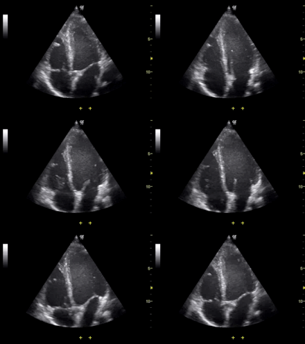
THEscan is a medical imaging technique that relies on the use of ultrasound, which makes it possible to “visualize” the inside of the body. Ultrasound of the heart, or echocardiography, allows you to observe all the structures of the heart, namely the valves and cavities (atria and ventricles). It also provides an overview of the heart muscle tone.
Why get an ultrasound of the heart?
Ultrasound is a painless and non-invasive examination: it is therefore prescribed as the first line when the doctor suspects a abnormality in the heart, for example after auscultation with a stethoscope.
The doctor can prescribe this examination in several situations, in particular for:
- Confirm the cardiac origin of symptoms such as shortness of breath, chest pain or discomfort
- Evaluate the impact of a disease (such ashigh blood pressure or pulmonary arterial hypertension) or certain medicines for the heart
- Diagnose a heart defect or disease of the heart muscle (cardiomyopathy)
- Diagnose a Heart Failure
- Look for or evaluate an anomaly of the heart valves (valvulopathy), these small “valves” which make it possible to prevent the blood from flowing back when it enters one of the cardiac chambers or that it is expelled by the heart
- Look for the presence of a clot
- Monitor the progress, and estimate the severity, of a previously diagnosed heart condition
- Study the pericardium (the envelope that surrounds the heart) or the thoracic aorta
Ultrasound of the heart is often coupled with a Doppler, a technique also based on ultrasound which makes it possible to visualize at the same time the blood flow in the heart and at the level of the valves and coronary arteries.
The exam
Ultrasound consists in exposing the tissues or organs that one wishes to observe to ultrasonic waves. It does not require any preparation.
The heart ultrasound, which lasts 10 to 30 minutes, can be done in two ways:
- Par transthoracic route : the probe is placed on the thorax (the patient must be shirtless), after the application to the skin of a gel facilitating the propagation of ultrasound. You will be lying down, but the doctor may ask you to turn on your back or on your side to better visualize certain structures.
- Par transesophageal route : the probe is associated with an endoscope (flexible tube) and inserted into the esophagus, to get closer to the heart and obtain better, more precise images. This examination is a little unpleasant because the probe must be introduced by the mouth (after local anesthesia and sedation). The transesophageal route is also used in people with overweight or having a lung disease which may interfere with the quality of the transthoracic image.
The doctor may also perform a so-called “stress” ultrasound of the heart, which consists of comparing the contractions of the heart muscle to the repos and during physical exertion.
A pharmacological stress test (triggered by a drug) may be done in patients who cannot exercise.
What results can we expect from an ultrasound of the heart?
Your doctor will inform you of the results of the ultrasound or cardiac Doppler ultrasound. In the event of an abnormality, other examinations (MRI, scanner, electrocardiogram, etc.) will probably be prescribed for a more in-depth cardiac assessment.
Depending on the situation, drug or surgical treatment may be prescribed, and appropriate monitoring will be put in place.
Read also : Our factsheet on arterial hypertension What is heart failure? All you need to know about blood clots |


