


Contents
Cattle often suffer from skin diseases. And these are not lichen, although there are enough of them. Various bumps and edema in cows are found in viral diseases and inflammatory processes. It could even be cancerous. A lump in the neck or head area of a calf may be a relatively harmless abscess or a serious fungal infection. There are many options when a cow has an incomprehensible swelling on the body.
Causes of bumps in a calf or cow
A bump is a loose concept. This word denotes both small solid formations with clear boundaries, and soft swellings, smoothly fading to “no”. There are many reasons for the appearance of certain “bumps”:
- allergy to parasite bites;
- inflammatory reaction to injection;
- actinomycosis;
- hypodermatosis;
- lumpy dermatitis;
- abscess;
- swollen lymph nodes in infectious diseases.
Sometimes the cause is determined independently if the appearance of the cones is very characteristic. But more often you have to call the veterinarian.
Allergic reaction
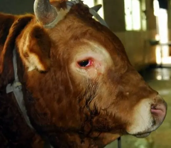
The first cases of the disease are recorded in calves. Allergy manifestations in cows are just as different as in humans. It depends on the individual characteristics of the calves. Food is manifested as swelling on the neck of the cow, and bumps all over the body. The latter go away on their own after the elimination of the allergen. Edema is more dangerous, since with its further development the calf may die from suffocation. Also, an allergic reaction in cows is expressed in lacrimation and abundant discharge from the nasal cavity.
The only really working way to treat the disease is to exclude the allergen from the environment. Without this, all other actions will be useless. Since it is difficult to find an allergen even in humans, calves with manifestations of the disease are usually handed over for meat. Antihistamines are prescribed by a veterinarian. He also determines the dose for the calf, based on its weight and age. Not all “human” antihistamines are suitable for cows. Some of them just don’t work, others can even kill the calf.
Provided that the lump arose at the injection site. Otherwise, with a high degree of probability – this is an abscess.
It rarely comes to bumps all over the body in calves and adult animals, this requires thin, delicate skin, but other signs of allergies are quite common.
Actinomycosis
A fungal disease that cows are most susceptible to. The name of the causative agent is Actinomyces bovis. Belongs to the genus Actinomyces. The opinion that this is a fungus is present in -language sources. English speakers indicate that it is a gram-positive rod-shaped bacterium. Pathogenic is an anaerobic type of microorganism.
The causative agent of the disease is not resistant to high temperatures: it dies within 5 minutes at 70-90 °C. But at sub-zero temperatures, the bacterium remains viable for 1-2 years. In 3% formaldehyde, it dies after 5-7 minutes.
Cases of infection are recorded year-round, but most often the disease of calves with actinomycosis occurs in winter and spring due to a decrease in immunity. The pathogen enters the body of a cow through any damage to the external integument:
- wounds of the oral mucosa or skin;
- cracks in the teats of the udder;
- castration wounds;
- when changing teeth in calves.
A distinctive feature of the disease is a dense bump (actinoma) on the cheekbone of a calf or an adult cow, since the bacterium most often affects the bones and tissues of the lower jaw.
When ripe, the cone opens, and creamy pus begins to come out of the fistula. With the development of the disease, an admixture of blood and pieces of dead tissue appear in the pus. The general body temperature of the calf is usually normal. An increase occurs only when the disease is complicated by a secondary infection or the bacteria spread throughout the body. Animals lose weight if the bumps “grew” in the pharynx or larynx. Tumors make it difficult for the calf to breathe and swallow food. Self-healing is very rare.
Treatment
An iodine solution is used intravenously. In the treatment of the disease, penicillin is used, which is injected into a bump on the cheek of a cow in a course of 4-5 days. Oxytetracycline has proven itself well. The dose for calves up to a year is 200 thousand units in 5-10 ml of saline. For animals older than 1 year, the dose is 400 thousand units. First, the antibiotic is injected into the healthy tissue around the calf’s cheek bump. Further, pus is sucked out of the fistula with a syringe and “replaced” with oxytetracycline. Course 2 weeks. Broad-spectrum antibiotics are also recommended. In advanced cases, they resort to surgical intervention and cut out the bump entirely.
Prevention
Calves do not graze on swampy pastures. Avoid giving roughage, especially with thorny plants, or steam it before distribution. The straw is calcined.
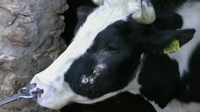
The characteristic location of the bump in a cow with actinomycosis
Hypodermatosis
A parasitic disease caused by gadflies from the genus Hypoderma. In common parlance they are called subcutaneous. The most common types:
- Hypoderma bovis;
- Hypoderma lineatum;
- Hypoderma tarandi.
The latter species is also called the deer gadfly. He lives in the northern regions and attacks mainly deer. The first two are subcutaneous gadflies of cattle, but bovis is a European species, and the lineatum is North American.
The genus Hypoderma includes 6 species. Parasites are not specialized. The same species lays eggs on any mammal that comes along, including cats and dogs. But they prefer large animals. Gadfly eggs are laid on the legs of cattle. The breeding season for parasites is from June to October. Each female lays up to 800 eggs, from which the larvae emerge after a few days.
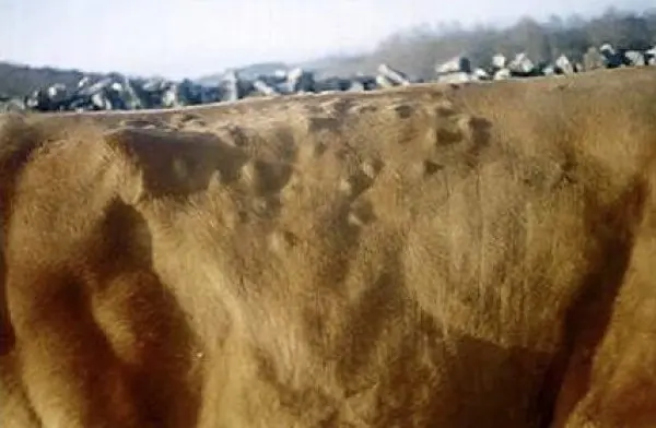
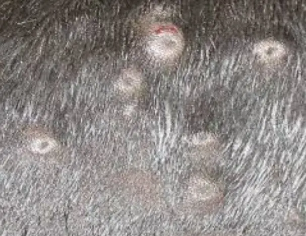
The latter penetrate the skin and begin to move upward. The final point of the “journey” is the back and sacrum of the cow. The move lasts 7-10 months. This duration of the disease is already considered chronic. The larvae of the last stage form hard cones with a respiratory hole in the middle on the upper line of the animal body. You can feel the nodules in the period from February to July. The larvae live in cones for 30-80 days, after which they leave the host.
The death of animals is not beneficial for parasites, but during the course of hypodermatosis, cattle lose weight, cows reduce milk yield, and calves slow down in development. After the larvae emerge and the holes in the cones close up, scars remain on the skin of the cow. This reduces the quality of the skins. The timing of slaughter is disrupted, since sick calves are not recommended to be slaughtered due to too much meat loss. Cones at slaughter must be cut. So lost up to 10 kg of meat.
Treatment and prevention
Preventive treatment is carried out in September-November. Use drugs that cause the death of the larvae of the first stage. Further, to prevent the spread of the disease next year, the herd is inspected in March-May. Check all the cattle grazing last summer.
It is best to feel the cow when examining. So there are more chances to find bumps in winter wool. Although usually the larvae “prefer” the back and sacrum, you can find nodules in other places. If, during a spring examination, a lump was found on the cow’s neck, this may also be a gadfly larva.
If nodules with breathing holes are found on animals, you should contact your veterinarian. He will prescribe drugs that destroy the larvae in the last stage and advise after what time it is possible to eat products from treated cows. With a strong infection of parasites from the cones will have to be removed manually to avoid intoxication of the body after the death of the larvae
In the end, the larvae from the cones will come out on their own, but before that they will severely exhaust their prey
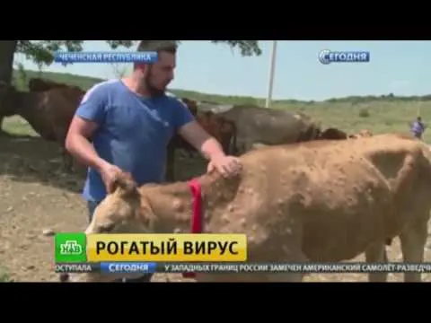
Lumpy dermatitis
A new viral disease “originally” from southern countries. Widespread in Africa and India. The main symptom is flat bumps all over the body of a calf or cow. The disease is caused by viruses related to goatpox. Both calves and adults are equally infected. The main carriers of lumpy skin disease in Our Country are blood-sucking insects. It is believed that in the southern countries the causative agent of the disease is carried by birds, in particular herons.
The loss of livestock is only 10% of the diseased animals. But dermatitis causes significant economic damage:
- a sharp decrease in the quantity and quality of milk;
- weight loss in calves fed for meat;
- abortions, infertility and stillbirth in breeding queens;
- temporary sterility of bulls.
The first sign of the disease is the appearance of dry bumps. And in any place, from the head to the udder and legs. The disease is little studied. Perhaps the location of the bump depends on the site of the initial entry of the virus.
If no action is taken, the bumps will very quickly cover the entire body of the cow, forming a kind of hard coating instead of skin. The rapid spread is due to the fact that the virus is carried through the bloodstream.
Symptoms of Lumpy Dermatitis
The latent period of the disease in natural conditions in cows lasts from 2 to 4 weeks. In the acute form of lumpy dermatitis, the following are characteristic:
- temperature 40 °C for 4-14 days;
- lacrimation;
- refusal to feed;
- mucus or pus from the mouth and nose;
- the appearance of cones 2 days after the transition of dermatitis to the clinical stage;
- the appearance of nodules throughout the body.
In severe cases of the disease, bumps appear on the mucous membranes of the oral and nasal cavities, vulva and foreskin. They also often occur on the eyelids, scratching the cornea. Due to constant irritation, the cornea becomes cloudy and the cow goes blind.
Usually bumps of lumpy dermatitis have a diameter of 0,2-7 cm. They are round in shape, clearly defined. In the center of each bump there is a depression that turns into a “cork” after 1-3 weeks. Later, the tubercle opens. It oozes a foul-smelling mucus.
After recovery, the bumps disappear. Where they were, hair falls out and skin flakes off.
Later, they dissolve or turn into dry scabs, under which there is granulation tissue.
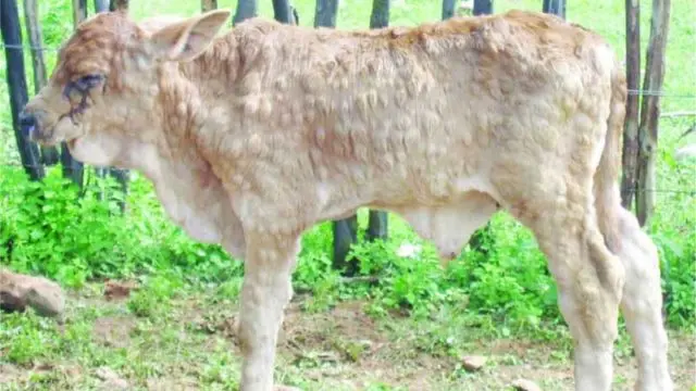
Calf with advanced form of lumpy dermatitis
Treatment and prevention
Neither one nor the other in application to lumpy dermatitis does not exist. Treatment of calves is carried out symptomatic, treating festering wounds with disinfectants. A course of antibiotics is given to cows to prevent the development of a secondary infection that penetrates through damaged skin.
As a prophylaxis of the disease, a live goatpox vaccine is used. But this does not always work. There is no way to passively prevent the disease.
Close-up of dermatitis bumps showing hollows in the middle of the tubercles, which later turn into separating plugs
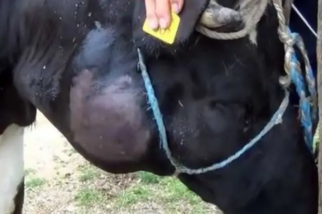
Abscess
Abscesses are common in cows and calves. Most often they occur due to injuries of the mucous membranes when eating roughage. It is also possible that inflammation occurs when the skin is damaged. Sometimes it is a reaction after vaccination. Practice shows that a hard hot bump on the neck of a cow is an abscess in the initial stage. While the abscess matures or is deep, the bump is solid. As the abscess matures, the tissues become soft. At any stage, the tumor is painful.
If the pus “went” to the outside, the skin at the site of the abscess becomes inflamed, the hair comes out. But abscesses located close to internal cavities often break through. The latter is especially dangerous for calves, since the tumor is very large and blocks the airways, and the animal can choke with a burst of purulent mass.
With the “internal” opening of suppuration, the inflammatory process often passes into a chronic stage. A capsule forms around the focus of inflammation, and the abscess lump looks hard from the outside.
The treatment is no different. They wait until the abscess matures, and open it, giving pus an outlet.
The vacated cavity is washed with disinfectant compounds until the solution begins to pour out clean. It is undesirable to sew up the wound, as drainage is necessary. Dead tissue comes out for a few more days. In addition, the cavity must be washed every day. And sometimes several times a day.
An abscess on the cheeks of calves and cows is often caused by damage to internal tissues when changing or improper grinding of teeth.
What to do if a cow or calf has bumps on its neck
First of all, find out the cause of the appearance, since the method of treating cones depends on the type of disease. The abscess is often heated to hasten its “ripening” and open. A bump on the jaw of a cow may be an inflamed lymph node: a symptom, not the cause of the disease. And even with the “simplest” case, the defeat of the animal by gadfly larvae, you will have to call a veterinarian. Without surgical skills, it is better not to open the bumps on your own.
The only option when hardly anything can be done is a bump after vaccination. Worst of all, animals react to anthrax. After this vaccine, bumps or swelling often occur at the injection site.
Conclusion
If a calf has a lump on its head or neck, the cause of its appearance is first determined. Since it is unlikely to be possible to do this on your own, you need to invite a veterinarian. In some cases, the treatment of “bumps” must be started as soon as possible.