


Contents
In line with its mission, the Editorial Board of MedTvoiLokony makes every effort to provide reliable medical content supported by the latest scientific knowledge. The additional flag “Checked Content” indicates that the article has been reviewed by or written directly by a physician. This two-step verification: a medical journalist and a doctor allows us to provide the highest quality content in line with current medical knowledge.
Our commitment in this area has been appreciated, among others, by by the Association of Journalists for Health, which awarded the Editorial Board of MedTvoiLokony with the honorary title of the Great Educator.
A chromosome is a collection of a given number and type of genes, inherited from both parents. A normal human chromosome consists of 23 pairs of chromosomes, and any changes in the number or mutation of genes can lead to serious diseases.
Chromosome – what is it?
A chromosome is the thread-like structure responsible for transmitting genetic information. Thus, it is a platform for gene transfer, conditioning the development of an organism and its mode of action, as well as personality and appearance traits.
Where is the chromosome? In eukaryotes, including humans, the chromosome is located in the cell nucleus. A healthy cell has 46 chromosomes in pairs. 22 pairs make up the autosomes, while the 23rd pair consists of sex chromosomes (allosomes).
A human inherits one of the chromosomes in a pair from the father and one from the mother. The definition of a chromosome defines it as the set of the basic units of heredity, or genes.
What is a chromosome made of?
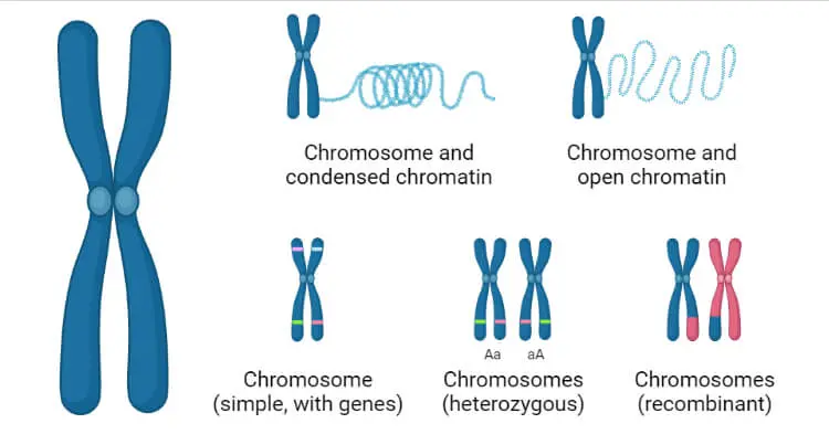
The structure of the chromosome is very simple, although it varies depending on the stage of cell division. The chromosome is made up of two arms which are maximally shortened in the prophase. In subsequent phases, it lengthens until it finally forms a shape known from descriptions and figures – its arms form twin chromatids that connect at a point called a centromere. This is how you can see the chromosome in the metaphase stagethat is, just before cell division.
It is also important what it contains. The human chromosome consists of:
- deoxyribonucleic acid, or DNA – a thin and long molecule that is a collection of genes, and thus information on how to code individual amino acids; DNA from one chromosome would be several centimeters long when stretched,
- histones, i.e. proteins constituting the structure around which DNA is wound and condensed,
- organizer of the nucleolus, i.e. secondary constriction (in acrocentric chromosomes),
- satellites, or trabant (not always).
The structure in which double-stranded DNA is wound onto histone protein molecules is called chromatin. Thanks to it, the chromosome is a safe hiding place for genetic material.
Find out more: “Certain diseases caused by chromosomal abnormalities”
Chromosome – types
There are several types of chromosomes due to their structure.
- The acrocentric chromosome. Its characteristic feature is one short arm in which in the secondary constriction (nucleolus organizer) there are sequences coding for nucleologenic areas. The short ‘p’ arm on the acrocentric chromosome places the centromere almost at the end of the structure. The acrocentric chromosomes include chromosomes 13, 14, 15, 21, and 22.
- Metacentric chromosome. A type of chromosome where the midweb is exactly on the middle of the chromosome. The arms form the letter V in it and are of equal length.
- Submetacentric chromosome is one in which the centromere is permanently located near, but not exactly at, the center of the chromosome. Thus, in the process of metaphase and anaphase, it has the form of the letter L.
- The telocentric chromosome. In this type, the chromosome has one arm and the centromere is at the end of it. It does not occur in humans.
Homologous chromosome – what is it?
The human chromosome consists of pairs that are called homologous chromosomes. They are almost identical – they have the same shape and size, and have similar genes (genetic information). However, they can exist in a different form – alleles. Then we are talking about heterozygote. However, if the form of the gene is the same, the cell is called homozygous. Homogeneous chromosomes make up autosomes, or non-sex chromosomes.
Homologous chromosomes form a pair of chromosomes, one from the mother and the other from the father. Pairing occurs for a short time during meiosis, then the homologous chromosomes diverge to the opposite poles of the dividing cell. This is how a gamete is formed, composed of cells with a haploid number of chromosomes.
Also read: «What do our genes say about us?»
Sex chromosomes
Next to the 22 pairs of autosomes, there is a 23rd pair of allosomes in the chromosome. The allosome is the sex-determining chromosome. Both men and women have the same X chromosome, but the other is different.
The X chromosome is always inherited from the mother. In women, the second X chromosome from the father determines the female sex. So the girl will have an XX chromosome pattern. The Y chromosome, in turn, determines the male sex. In men, again one (X) chromosome is from the mother and the other (Y) from the father, making a total of a pair of XY chromosomes.
The inheritance of the Y or X chromosome from the father does not occur when the parents’ reproductive cells (gametes) are mergedbut still in the process of meiosis in these cells. During this division, the X or Y chromosome goes to the gamete individually. Compared to other cells of the body, these reproductive cells have 22 autosomes and only one chromosome – X in women, X or Y in men.
Chromosome aberration can occur during the formation of a zygote with a specific karyotype. In such cases, in addition to the 46 standard chromosomes, an additional allosome appears or the structure of the chromosomes is modified.
- Chapel syndrome (also male XX syndrome) describes a male with the 23rd pair of chromosomes typical of women. It is defined as an intersex disorder in which, in most cases, the SRY gene is transferred from the Y chromosome to the father’s X chromosome during meiosis. The paternal gamete with the X chromosome combined with the maternal gamete genetically produces the XX allosomes, or the female chromosomes, but in fact the SRY gene gives the baby male characteristics.
- Androgen insensitivity syndrome applies to people who, in terms of second and third-order traits, are female, but genetically male, and therefore female, who have male XY chromosomes. There are two levels of this disorder. In the first one, the female genitalia is fully developed, but the testicles are located in the abdominal cavity or in the inguinal canal, around which no further elements of the male genitalia have developed. The testes were formed by the SRY gene located on the Y chromosome. However, the male hormones they synthesize do not act on cells. In turn, in the total insensitivity to androgens in the body, there are no traces of male genitalia. There are also no periods.
- Turner syndrome affects women with only one sex chromosome, known as the X0 karyotype. The absence of a second X chromosome may be complete or partial. In this case, the features of the appearance are recognizable: short stature, not very expressive female features in the figure and face, a short, fluffy neck, clearly defined eyes, drooping eyelids, poor facial expressions. Often times, people with a single X chromosome suffer from infertility.
- Klinefelter syndrome affects men who have more than one X chromosome. The syndrome is characterized by the karyotype of 3 chromosomes in the 23rd pair, and sometimes even 4 or 5. People with this karyotype are sometimes diagnosed with mental retardation – from moderate to severe degree. In the case of XXY chromosomes, there are also high growth, slightly feminine figure, poorly developed musculature, long arms and legs, gynecomastia, no or delicate facial hair, small testicles, low libido and erectile dysfunction, often also a tendency to overweight. Combinations of XXXY and XXYY chromosomes are associated with much more severe mental retardation, as well as defects such as ocular hypertelorism and flat nasal bridge. In turn, the occurrence of the XXXXY karyotype, i.e. Fraccaro syndrome, is characterized by short stature, mongoid eyelid positioning (as in Down’s syndrome), severe mental retardation and hypotension.
- XYY Team, i.e. super male or supermen syndrome. It is characterized by 2 Y chromosomes in the karyotype. It is not associated with complications or diseases. However, with XYY chromosomes in men, there may be a lower IQ, learning and concentration difficulties.
- X chromosome trisomy means meta woman or super woman. A person with trisomy X is characterized by well-marked third-order female features, high stature and standard fertility, although in extreme cases menstrual disorders may occur. There is a slight disability and learning difficulties. There are also tetrasomy and pentasomy of the X chromosome, where there are 4 or 5 X chromosomes in succession.
Also read: “New genes responsible for autism and mental retardation discovered”
Chromosomes – diseases
Genetic diseases are the result of an abnormal structure or number of genes or chromosomes. Some of them are inherited, while others result from new gene mutations or changes in the structure of the chromosome as a result of improper distribution of autosomes in the parent’s gamete and, consequently, modification of their number in the child’s body. Genetic diseases are congenital and are diagnosed in utero or right after birth.
The most common diseases caused by a distorted gene or chromosome include:
- Down syndrome – chromosome 21 trisomy, i.e. the occurrence of an additional such chromosome in the karyotype, occurs once in 650-700 live births, often occurs in spontaneously aborted fetuses; children who survived the first year of life should live to 1-50 years, the male will be sterile, and the female will have a 60% probability of passing the defective chromosome to their offspring,
- Edwards’ syndrome – occurs due to an extra 18 chromosome in the karyotype, usually in transferred pregnancies in which low fetal mobility, placental underdevelopment or fetal underdevelopment has been detected,
- Patau’s syndrome – results from trisomy of chromosome 13 and is extremely rare, because its appearance usually leads to miscarriage, and the mortality rate of children under 1 year of age is close to 90%,
- Prader-Willi syndrome – a rare genetic disease resulting from a slight reduction of chromosome 15 obtained from the father, possible to be diagnosed after birth – is associated with specific features of the external appearance, but also with reduced muscle tone and behavioral disorders, including aggressive behavior,
- Angelman syndrome – also resulting from the loss of a certain fragment of chromosome 15, but inherited from the mother, slightly more common, called the happy puppet syndrome due to uncontrolled bouts of laughter,
- Williams syndrome – resulting from the deletion of chromosome 7, affects 1 in 20 births, manifests itself with a characteristic “elven” face, mild temper and slight impairment, often the so-called absolute hearing,
- Fragile X syndrome – a genetic disease caused by a dynamic mutation (duplication of a gene segment) in the FMR1 gene located on the X chromosome, activates more often in boys than in girls and is manifested by a reduction in the level of intellectual development (often similar to autism).
Read more about Angelman Syndrome and Prader-Willi Syndrome
Chromosome 21 – what is it responsible for?
One of the most famous autosomes in the human cell is chromosome 21. This is because of the genetic disease that an additional chromosome carries, Down syndrome. It is a relatively common condition compared to the frequency of other genetic diseases.
However, the chromosome in pair 21, when it is modified, can lead to other diseases.
- Alzheimer’s disease. Although there is no scientific evidence to support a specific cause of the disease, there are studies by John Hardy and David Allsop from 1991 hypothesizing that the cause may be a defect in the APP gene located on chromosome 21.
- Homocystinuria. A metabolic disease associated with over a dozen different mutations in the CBS gene.
- Amyotrophic lateral sclerosis. The genetic type of this disease is the result of a mutation in the SOD1 gene, which is found on chromosome 21.
Philadelphia chromosome
In professional literature, it appears under the English notation Philadelphia chromosome. It was discovered by two scientists – Peter Nowell (University of Pennsylvania, Philadelphia) and David Hungerford (Philadelphia Cancer Research Institute). In its role, the type of mutation occurring in the Philadelphia chromosome is important.
It is about translocation of the BCR gene from chromosome 22 towards the ABL gene on chromosome 9, as a result of which the latter is characterized by a longer arm. The ABL gene belongs to proto-oncological genes, i.e. genes that can lead to neoplastic processes. As a result of fusion with the BCR gene, it becomes active and becomes an oncological gene, influencing the excessive formation of white blood cells in the bone marrow.
The Philadelphia chromosome is detected in the case of a blood cancer, namely:
- chronic myeloid leukemias (occurs in 95% of cases),
- acute myeloid leukemias (more than 1% of cases),
- acute lymphoblastic leukemias (25-30% of cases in adults and 6% in children).