Contents

Chest deformities are curvature of the sternum and ribs, which can have varying degrees of severity.
Congenital deformity of the sternum is not common and is diagnosed in no more than 2% of people worldwide. Most often, acquired deformities are found in people, which develop in childhood and adolescence.
Congenital deformities occur due to genetic abnormalities. Acquired curvature of the thoracic region develop against the background of various diseases.
When the chest is deformed, the bones and muscles that protect the internal organs from external damage suffer. Treatment of such a pathology should be started as early as possible. If therapy is absent, the likelihood of severe complications increases. They relate to the work of the cardiovascular system, respiratory and digestive organs.
Causes of breast deformity
Congenital deformities of the chest can occur under the influence of the following factors:
Genetic predisposition.
Turner Syndrome, Down Syndrome, Marfan Syndrome.
Violations caused by the shaping of the skeleton: ribs, spine, chest.
Acquired deformity of the chest may be due to the following factors:
tuberculosis of the bones.
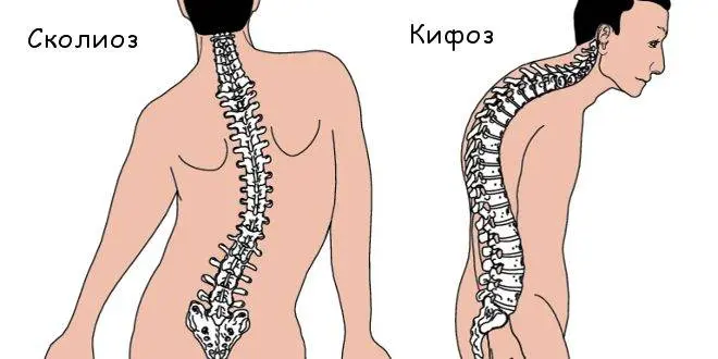
Scoliosis and kyphosis.
Injuries and burns of the chest.
Rickets.
Diseases of the lungs.
Symptoms
The main symptoms of chest deformity are:
Changes in the structure of the spine. Moreover, not only the thoracic, but also the cervical and lumbar regions will suffer.
Curvature of the pelvic bones, different lengths of the legs. These symptoms are more characteristic of scoliosis.
Increased muscle tone on one side of the body, the formation of a muscle roller.
Expansion of the intercostal spaces. In appearance, this area will resemble a hump, but it is located in front.
Violations in the work of the respiratory system and the heart.
Torsion – displacement and twisting of the vertebrae.
Curvature of the neck, irregular shape of the skull, facial asymmetry.
Pain in chest and back. It occurs due to the infringement of nerve fibers.
Classification and species

Chest deformities are divided into congenital and acquired. They, in turn, also have certain subspecies. There is a classification of deformations depending on the place of their localization and on the severity of the violation.
So, 2 large groups of chest deformities:
congenital deformities.
Acquired deformities.
Congenital deformities are divided into the following subspecies:
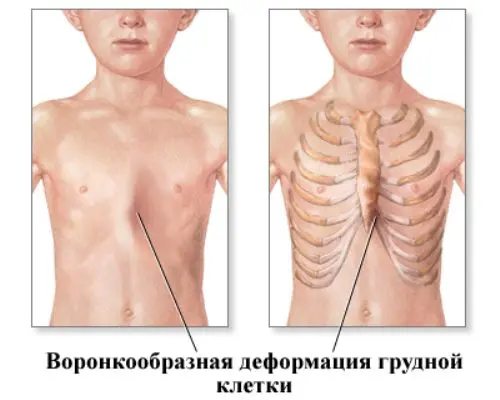
funnel deformity. At the same time, the chest in a person is sunken, it looks pressed inward. Experts call such a violation the cobbler’s chest.
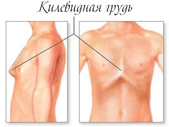
Keeled deformation. The patient’s chest is pushed forward and resembles a boat keel. In the people, such a violation is called “chicken breast”.
Flat. Both the chest and the ribs are flattened towards the anteroposterior axis.
There are congenital deformities that are diagnosed quite rarely.
These include:
Sternoskisis or congenital cleft sternum. In this disorder, the sternum may be partially or completely split.
Rib-muscular defect. With this violation, not only the chest suffers, but also the spinal column, as well as muscles and internal organs (Poland’s syndrome).
Arched chest or Currarino-Silverman syndrome. This deviation is not common.
Acquired deformities are classified as follows:
Emphysematous deformity that develops against the background of emphysema. A person’s chest in appearance begins to resemble a barrel.
Paralytic deformity, accompanied by an expansion of the space between the ribs. At the same time, the side of the chest becomes smaller in size, and the collarbone and shoulder blades begin to bulge. This type of violation leads to diseases of the lungs and pleura.
Navicular deformity. The patient has a rather long depression, which is shaped like a boat. This disorder is a consequence of syringomyelia.
Kyphoscoliotic deformity develops with a curvature of the spine, or after tuberculosis.
According to the form of deformation can be of the following types:
Symmetrical.
Asymmetrical.
Задние.
Front.
Side.
The severity of the deformity is determined by the depth of the chest displacement and the degree of pressure on the heart.
In this regard, there are:
Compensated deformity (first degree) with an impression of no more than 2 cm. With such a deformity, the heart does not suffer.
Subcompensated deformity (second degree). The depression is 2-4 cm. In this case, the heart can move by 3 cm.
Decompensated deformity (third degree). The depression exceeds 4 cm, the heart is displaced and reduced in size. This pathology is a health hazard.
In childhood, decompensated deformity is rarely diagnosed. The sooner corrective treatment is started, the better the results will be. Therapy should be carried out until such time as there are no complications from the heart and lungs.
congenital deformities
With congenital deformities, the shape of the chest will be distorted in a person. This is due to the underdevelopment of the ribs and the muscular frame. Sometimes babies don’t have ribs at all.
Funnel chest deformity
The funnel-shaped type of deformity of the sternum is more common than others. This disorder is diagnosed in 90% of all cases of dysplastic curvatures. Girls are affected less frequently than boys. “Shoemaker’s chest” is manifested by a pronounced depression in the center and in the lower part of the chest. This is due to the underdevelopment of the cartilage of the ribs.
Violation can be provoked by factors such as:
Improper growth of cartilage and bones.
Congenital defects of the diaphragm, which are accompanied by its shortening.
Incorrect position of the fetus in the uterus.
If the deformity is severe, the child will suffer from surges in blood pressure, from curvature of the spine. There are violations in the work of internal organs. First of all, the heart and lungs suffer.
Keeled deformity of the chest (chicken breast)
In a patient with a chicken breast, the ribs and sternum protrude forward. Often this pathology is diagnosed in preschool children. This violation does not affect health and is only a defect in appearance. To date, doctors do not know for certain the causes of such a violation, but there is an assumption that it may be associated with anomalies of the genetic plan.
Over time, this deformation will progress. However, it cannot cause serious harm to internal organs. However, the heart with such a violation may change shape and will resemble a drop. A person will begin to get tired faster, he may experience shortness of breath and tachycardia.
Flat chest
In a person with this disorder, the front and back of the sternum is smaller than in other people. It is impossible to call a flat chest a disease. Deformation refers to the features of the development of a particular organism. It does not affect the work of internal organs, so corrective measures are not indicated for such patients.
A person with a flat chest has the following body features:
High growth.
Too narrow shoulders.
Long limbs.
Mismatch of height and body weight.
Children with a flat chest are more likely to get cold infections, sometimes there is a developmental delay.
Rib-muscular defect (Poland’s syndrome)
Poland’s syndrome is a severe pathology of the development of the rib-muscular apparatus. The patient has a deformed chest and spine, which leads to changes in the internal organs. Bone structures are often displaced.
People with Poland syndrome have several disorders at once:
The pectoralis major and minor muscles are absent.
Fingers spliced or too short.
The ribs are either absent or severely deformed.
In half of the people with this defect, the deformities are caused by genetic abnormalities.
Arched sternum (“upper keel”, Currarino-Silverman syndrome)
Arched sternum is not often diagnosed. This violation is manifested by a protruding groove, which is located in the upper part of the chest. The costal cartilage will be overgrown. That part of the chest, which is located on the opposite side, will have a normal appearance. Such a deformation does not affect a person’s health, but spoils his appearance. To get rid of it, you will need the help of a plastic surgeon.
Congenital cleft sternum (sternoskisis)
With this violation, the sternum will be partially or completely split. This anomaly is considered rare, although it is diagnosed in about 2% of the total population of the Earth.
Splitting can be H-shaped, top or bottom. This pathology is considered dangerous, as it progresses all the time. The heart of a sick person will not be covered by the ribs, it is located directly under the skin. If you look at the chest, you can see the beating of the organ. All people with sternoskisis require surgery.
Acquired deformities
There are a wide variety of deformations that develop in a person over the course of life. They are caused by various diseases and in themselves worsen the health of the patient.
Emphysematous chest. This pathology is caused by emphysema of the lungs. The human breast takes on the shape of a barrel. The patient suffers from breathing problems, then heart disorders join.
Paralytic chest. This deformity develops due to pulmonary pathologies. The lateral parts of the sternum and its anteroposterior regions decrease, and the intercostal spaces become wider. On both sides, the sinking is different, therefore, during breathing, the shoulder blades move asynchronously.
Scaphoid chest. There is a recess in the center of the sternum. The scaphoid chest develops with pathologies of the central nervous system, when cavities form in the spinal cord and in the medulla oblongata. This disorder is called syringomyelia.
Kyphoscoliotic chest. Kyphoscoliotic chest is a consequence of bone tuberculosis. Sometimes the deformity develops against the background of other pathologies of the chest organs. In itself, this curvature negatively affects the work of the heart and lungs. The disorder is incurable.
Diagnostics

Without fail, the patient is prescribed a chest x-ray, which is performed in two projections with the calculation of the Gizhitsk index. This study allows you to determine the severity of the curvature, to detect whether there is a displacement of the heart, as well as disturbances in the functioning of the lungs. Scoliosis is diagnosed using x-rays.
Additional examination methods include:
Computed tomography. It is prescribed to patients with severe deformity, the study is carried out in order to clarify whether the patient needs the help of a surgeon. CT provides information about the compression of the heart, the degree of its displacement, and the condition of the lungs.
Magnetic resonance imaging. It is carried out with significant deformations and its asymmetric varieties of 3 or 4 degrees.
In order to assess the work of the heart and blood vessels, the patient is given spirography, ECG and ECHOCG.
Treatment of deformation changes in the chest

Features of therapy depend on the type of deformity. The doctor also takes into account at what stage of development it is. As soon as possible, treatment is started for patients in whom the work of the heart and lungs is disturbed due to deformation.
The main objectives to be achieved by therapeutic measures:
Get rid of deformation.
Eliminate pressure on the internal organs and bring their performance back to normal.
Eliminate the defect of appearance, improve the quality of human life.
Conservative treatment
It will not be possible to cope with the deformation by conservative methods. They are practiced only to stop the progression of the disease.
Methods of influence on the human body can be as follows:
Massage. Point methods are used, only a doctor can implement them.
Physiotherapy.
Wearing a corset.
Swimming.
Breathing exercises.
To cope with the deformity that has already occurred, the help of a surgeon is required.
vacuum bell
If the deformation has just begun to form, then a method called a vacuum bell is used. It allows you to cope with the violation, provided that the human chest is supple. A vacuum is created in it, which corrects the existing curvature. The maximum effect of the procedure can be achieved at the age of 6-7 years. If therapy fails, then surgery is required.
Ferre dynamic compression system
This system is designed to correct the existing asymmetry in the chest area. Each specific device is developed for a person individually.
It consists of several components:
A plate represented by an aluminum alloy. It is adjusted to the part of the chest that protrudes forward.
The mechanism responsible for supporting the torso. It is located on the back side.
A device for measuring the pressure exerted by a structure on the human body.
This system is used to correct deformities in childhood, as well as as part of a complex treatment regimen.
If the age of the patient is older than 20 years, then a significant result cannot be achieved. Such treatment is well tolerated by all patients, since it does not impair their quality of life and does not cause discomfort.
As defects are corrected, the structure is transformed by adjusting the applied pressure. The timing of treatment varies widely and depends on the specific clinical case. If the design was used in the early stages of the formation of curvature, then it becomes possible to avoid surgery.
Orthoses
Orthoses are medical devices that are actively used to correct chest deformities. When a patient is diagnosed with a keeled curvature, he is prescribed a corset that gives the upper body the correct position.
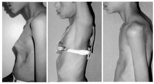
The area of the chest that protrudes forward will be squeezed by the corset. Its regular use allows you to eliminate the existing deformation, as well as correct the defect in appearance. During treatment, the doctor must adjust the pressure exerted by the orthosis.
Significant results can be achieved only when the treatment was started at an early age. Teenage deformities can be corrected, since in this period the bone tissue is quite flexible and responds to outside influences. With severe deformities, it will not be possible to achieve results only with the help of orthoses.
It will take a long time to wear corrective devices. The minimum time of their use is 12 hours a day. You need to tune in to long-term treatment. With this approach, after 1-1,5 years, the chest will acquire an anatomically correct position.
Surgical correction
It is possible to cope with chest deformity with the help of surgery, but it is important to choose the technique that suits a particular patient. There are many corrective operations, but most of them are reduced to osteotomy. During the procedure, the doctor performs a bone incision, after which he implements the necessary manipulations.
Sterno-chondroplasty. Mark Ravich method. An incision is made on the anterior chest wall, which crosses the chest across. The surgeon then separates the muscles, excised the costal cartilages and the sternum (sternotomy). The next step is setting up a correction plate. After that, the fabrics are sewn together. Such an intervention is very effective, but at the same time traumatic for the patient. Many people refuse the operation, since the incision is made on the chest, from its front side. Postoperative sutures do not look very aesthetically pleasing.
The Abramson method. This method of treatment is not as traumatic as the intervention described above. Therefore, this type of operation is chosen by a greater number of patients with deformity. The plates during its implementation are sutured to the ribs. When the desired effect is achieved, these plates are removed.
Kondrashin’s method. An incision is made on the anterior sternum. Make it not horizontal, but vertical. Then the deformed cartilage is excised using the wedge-shaped method. Deformed bones are excised in a similar way, the xiphoid process is crossed. The operation ends with stitching the dissected tissues and suturing the chest.
The Tymoshenko method. If the operation is performed on a boy, then one incision is made under the pectoral muscles. When a girl needs correction, two incisions are made. In the area of deformation, the skin is exfoliated, the muscles are separated from the ribs, and the ribs themselves are removed while preserving the perichondrium. The rib is shortened by a few centimeters. In the upper part of the deformation, a metal plate is attached, which is fixed on the ribs. It repeats the anatomically correct shape of the chest. A few months later, this plate is removed.
Nass method. This operation is indicated for pectus excavatum. It is characterized by low trauma. During the procedure, two incisions are made on the sides of the chest. Metal plates are inserted into them, which give the sternum the correct shape. A few years later, these structures are removed. The procedure is performed under general anesthesia. The patient must remain in the hospital for 7 days. Then for about 30 days he will need to limit physical activity, only walking is allowed. Sports activities are excluded for the next 3 months. In six months, a person will be able to return to a full life, as rehabilitation will be completed. He ceases to feel the plates inside the chest, the pain goes away.
Video: health “Shoemaker’s chest” – chest deformity:


