Contents
Cerebral aneurysm – what is it?
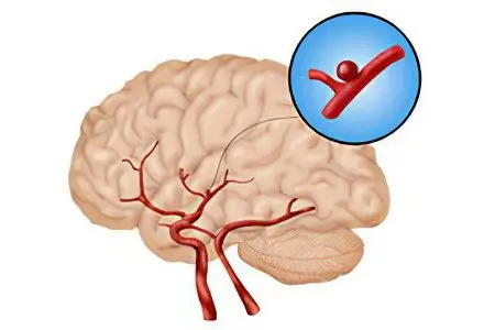
A cerebral aneurysm is an enlargement of one or more blood vessels in the brain. This condition is always associated with high risks of death or disability of the patient if an aneurysm ruptures. In fact, an aneurysm is a protrusion of the vascular wall that occurs in a particular part of the brain. Aneurysms can be congenital or develop over a lifetime. At the same time, it damages the integrity of blood vessels and often leads to cerebral hemorrhages. They are the main threat not only to health, but also to human life. As a rule, an aneurysm rupture occurs in people aged 40-60 years.
Since the diagnosis of cerebral aneurysm is associated with certain difficulties, it is rather difficult to determine the real degree of its prevalence among the population. However, the statistics are that for every 100 people, 000-10 of them have an aneurysm. Post-mortem autopsies show that aneurysms that did not provoke a rupture of a cerebral artery in 12% were not diagnosed during a person’s lifetime. They are discovered by chance, as they do not give any symptoms.
Nevertheless, the leading threat of an aneurysm has been and remains a rupture of the vessel with hemorrhage in the brain. This situation requires urgent medical attention, which is not always effective. The harsh statistics is such that against the background of subarachnoid bleeding, 10% of patients die almost instantly, even before doctors have the opportunity to provide them with first aid. Another 25% of people die on the first day, and up to 1% die within the first three months after a cerebral hemorrhage occurs. Summing up the sad result, we can say that the frequency of death due to rupture of an aneurysm of cerebral vessels is equal to 49%. Moreover, the death of patients occurs more often in the first hours or days after the cerebral catastrophe has occurred.
Despite the high development of medical science, the only method of treating cerebral aneurysms is surgery. However, even it does not provide 100% protection against death. However, the risk of death from a sudden aneurysm rupture remains 2–2,5 times higher than the risk of death during or after surgery.
As for the countries where cerebral aneurysms are most common, the leaders in this regard are Japan and Finland. If we turn to gender, then men suffer from this pathology 1,5 times less often. Women are three times more likely to have giant protrusions. Aneurysms are very dangerous for women in position.
What causes a cerebral aneurysm to form?

The leading cause of the formation of an aneurysm can be called a violation of the structure of any layer of the vascular wall, of which there are three: intima, media and adventitia. If these three shells are not damaged, then an aneurysm will never form in them.
The reasons that provoke its formation include:
Transferred inflammation of the membranes of the brain – meningitis. Against the background of the disease itself, it can be quite difficult to identify the symptoms of an aneurysm, since the person’s condition remains difficult. After meningitis is treated, defects can remain on the walls of cerebral vessels, which will later lead to the formation of an aneurysm.
Head injuries that provoke stratification of the vascular walls.
The presence of a systemic disease. The danger is bacterial endocarditis, untreated syphilis and other infections that reach the vessels of the brain with blood flow and damage them from the inside.
Some congenital diseases (Marfan syndrome, tuberculous sclerosis, Ehlers-Danlos syndrome, systemic lupus erythematosus, congenital polycystic kidney disease, and some others).
Hypertension.
Autoimmune diseases that cause damage to the arteries.
Atherosclerosis.
Other causes, including: cerebral amyloid angiopathy, malignant tumors, which will not necessarily be localized in the brain.
Cerebral aneurysm is not inherited, however, it can occur against the background of diseases to which a person has a predisposition. Such diseases, for example, include hypertension, atherosclerosis, some immune and genetic pathologies.
What is a cerebral aneurysm?
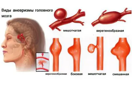
There are several types of classification of cerebral aneurysms, each of which has its own classification criterion. Having determined what kind of aneurysm the patient has, it is possible to choose an effective treatment and make the most accurate prognosis.
Types of vascular aneurysms depending on their shape.
The aneurysm is saccular. This aneurysm is more common than others, if we consider exclusively the vessels of the brain.
Fusiform aneurysm. It most often forms on the aorta, but rarely develops in the brain. The aneurysm has a cylindrical shape and causes a fairly uniform expansion of the vascular wall.
The aneurysm is exfoliating. It has an oblong shape and is located between the layers that make up the wall of the vessels. Most often, such an aneurysm also occurs on the aorta, which is explained by the mechanism of its formation. It is formed when there is a defect in the intima, where blood gradually begins to enter. This leads to delamination of the wall and the formation of a cavity. In the vessels of the brain, the blood pressure is not as high as in the aorta, so this type of aneurysm is rarely found here.
Types of vascular aneurysms depending on their size. The smaller the aneurysm, the more difficult it is to detect it during diagnostic measures. In addition, such aneurysms do not give severe symptoms. Large aneurysms, in turn, put pressure on brain structures and cause related symptoms. Don’t assume that small aneurysms aren’t dangerous, as they all grow over time. How quickly the aneurysm will increase in size is unknown.
Large aneurysms are those larger than 25 mm.
Aneurysms are medium – their size is less than 25 mm.
Small aneurysms are those whose diameter does not exceed 11 mm.
Types of vascular aneurysms depending on their location. This criterion largely determines the symptoms of the disease, since each segment of the brain is responsible for certain functions. So, a person may suffer to a greater extent from hearing, speech, vision, coordination, breathing, heart function, etc. The names of the types of aneurysm in this case come from the vessel on which it is located. In this regard, there are:
Aneurysms of the basilar artery (occurs in 4% of all patients).
Aneurysms of the posterior (26%), middle (25%) or anterior (45%) cerebral artery.
Aneurysms of the inferior and superior cerebellar arteries.
Depending on when the aneurysm formed, congenital and acquired defects are distinguished. Acquired aneurysms are more prone to rupture due to their high growth rate. Therefore, during the diagnosis, it is highly desirable to determine the time of formation of the protrusion. So, some aneurysms form in just a few days and quickly rupture. Other aneurysms, on the contrary, can exist for years and do not give themselves away.
Depending on the number of aneurysms, multiple and single formations are distinguished. Most often, a single protrusion is found in the brain – in 85% of cases. Risk factors for the formation of multiple aneurysms are serious brain injuries, or surgical intervention on its structures (we are talking about global operations), as well as congenital diseases that disrupt the quality of connective tissue. Naturally, the more formations a person has, the worse the prognosis.
What is a saccular aneurysm?
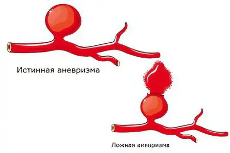
The reasons for the formation of saccular aneurysms most often come down to point damage to the vessel, or rather, one of its layers. As a result, the wall of the vessel begins to gradually bulge outward, which leads to the appearance of a sac that is filled with blood. Its bottom is most often wider than the hole through which blood enters.
In the presence of a saccular aneurysm, there is a risk of developing the following disorders:
Deterioration of the supply of certain sections of the artery with blood, due to its slower current.
Swirls of blood during its movement through a vessel with an aneurysm.
The presence of swirls leads to an increased risk of blood clots.
The risk of rupture of the vessel wall increases, as it turns out to be too stretched.
The brain can suffer due to compression of its tissues by an aneurysm, which increases in size.
Saccular aneurysms are also more likely to rupture and provoke the formation of blood clots when compared with other types of aneurysms.
What is a false aneurysm?
False aneurysms are not widespread, but they can occur. The defect is not a protrusion of the vessel, its damage in the form of a rupture. Blood through the existing damage in the vessel wall flows out of it and begins to accumulate nearby, forming a hematoma. When the damage is not epithelialized, and the leaked blood itself does not spread, then a cavity is formed in the brain tissues, connected to the vessel. Such an aneurysm leads to disruption of blood flow, but it is not limited by the vascular wall. Therefore, doctors prefer to call such formations pulsating hematomas.
At the same time, a person remains at risk of developing massive bleeding in the brain tissue, because the damaged vessel wall remains broken. As for the signs of a false aneurysm, it can manifest itself as a true aneurysm or have symptoms of a hemorrhagic stroke. Making a differential diagnosis is very difficult, especially in the early stages of hematoma formation.
What is a congenital aneurysm?
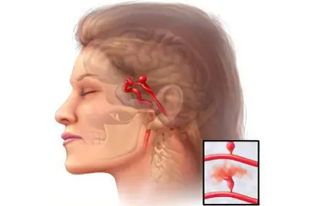
If he talks about congenital aneurysms, then they mean those that a person had at the time of his birth. They began to form during the intrauterine life of the fetus and do not disappear after birth.
The following reasons may lead to their formation:
Diseases transferred by a pregnant woman (viral infections are a danger in this regard).
The presence of a genetic disease that has a destructive effect on the connective tissue.
Intoxication of the woman’s body during gestation.
The presence of chronic diseases in a pregnant woman.
The impact of radioactive radiation on a pregnant woman.
Congenital aneurysms are most often found in those children whose mothers have undergone any harmful influence on the body from the outside. It is possible that the child will be born with other malformations, which happens very often.
It is quite difficult to make a single prognosis for each child with cerebral aneurysm. However, if the aneurysms are not false and the child has no other malformations, then the prognosis can be considered favorable, since the risk of rupture of congenital aneurysms is not high (their walls are quite thick). Nevertheless, a child from birth should be registered with a pediatric neurologist, since the presence of such an education in the brain can affect its development. If we consider the most severe cases, then congenital aneurysms are very large and sometimes incompatible with the life of the fetus.
How does a cerebral aneurysm manifest?

For a long period of time, an aneurysm of cerebral vessels may not give itself away. The protrusions rarely reach large sizes and form on small arteries (in the brain, all vessels are small). Therefore, the weak pressure that an aneurysm exerts on brain tissue is often not enough for a person to show any symptoms of the disease.
However, sometimes the course of the disease can be quite severe, which occurs in the following situations:
The aneurysm is large and puts a lot of pressure on parts of the brain;
An aneurysm is located in a place in the brain that is responsible for extremely important functions;
An aneurysm ruptures as a result of increased physical exertion on the body, against the background of stress, etc .;
Against the background of hypertension and other chronic diseases, aneurysm can give more pronounced symptoms;
Aggravates the course of the disease arteriovenous anastomosis.
Symptoms that indicate the presence of an aneurysm include the following:
Headaches that occur at different intervals and have different intensity.
Insomnia or increased drowsiness.
Nausea and vomiting.
Meningeal symptoms that may occur with aneurysms located in close proximity to the membranes of the brain.
Convulsions.
Deterioration of skin sensitivity, impaired vision, coordination, hearing. Specific manifestations of the disease primarily depend on where exactly the aneurysm is located.
Disturbances in the work of the cranial nerves responsible for the movement of small muscles. The patient may develop facial asymmetry, hoarseness, drooping eyelids, etc.
Possible consequences of cerebral aneurysm

Almost any symptoms of this pathology can be attributed to complications of cerebral aneurysm, since they all lead to certain disorders. So, it is difficult not to call loss of vision or hearing a complication, which is provoked by compression of the nervous tissue by dilated blood vessels.
In addition, an aneurysm can cause other dangerous consequences for human health, for example, which occur when it ruptures. Other complications occur somewhat less frequently, but carry no less threat.
Complications that can occur against the background of the presence of cerebral aneurysm:
Coma. If an aneurysm is formed in those parts of the brain that are responsible for the vital functions of a person, then he can fall into a coma. The duration of the coma can be different, and often lifelong. Moreover, despite high-quality and timely medical care, many patients never get out of this life-threatening condition.
Thrombus formation. In the cavity of the formed aneurysm, slowing down and disruption of blood flow can occur, which leads to the appearance of a blood clot. Most often, such a complication develops against the background of the presence of a large aneurysm. The location of the thrombus may vary: sometimes it occurs in the cavity of the aneurysm itself, and sometimes it breaks off and blocks the blood flow in smaller vessels. The more massive the thrombus, the more serious the threat to a person’s life, since in such a development of events he always suffers an ischemic stroke. However, with the provision of timely medical care, the life of the patient can be saved. Often, a blood clot can be dissolved with the help of drugs.
Formation of AVM. AVM is an arteriovenous malformation, which is essentially a defect in the vascular wall. This violation leads to partial adhesion of the vein and artery. The pressure in the cavity of the artery begins to fall, and some of the blood passes into the vein. This leads to an increase in pressure in the vein, and those areas of the brain that were fed from the artery begin to experience hypoxia. The AVM is indicated by the same signs that occur against the background of ischemic stroke. Sometimes the symptoms of AVMs are difficult to distinguish from the symptoms of a cerebral aneurysm. The larger the aneurysm, the more the vessel stretches, which means the higher the risk of AVM formation. With the development of this complication, surgical intervention is required.
Due to the fact that an aneurysm can provoke serious complications that threaten a person’s life, doctors, when it is detected, insist on performing an operation. Moreover, the need for surgical intervention is also due to the severity of the symptoms of the aneurysm itself.
Sequelae of ruptured aneurysm
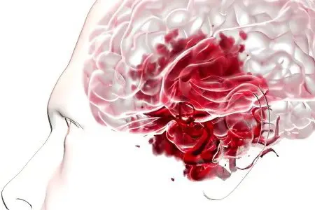
There are certain factors that can make a brain aneurysm more likely to rupture, including:
Experienced stressful situation;
Excessive physical stress on the body;
Hypertension or jumps in blood pressure;
The use of alcoholic beverages;
Infectious diseases occurring against the background of high body temperature.
After an aneurysm rupture has occurred, symptoms begin to increase sharply in a person, which in general is not characteristic of this disease. The patient’s condition deteriorates sharply and requires immediate medical attention. Signs that may indicate a ruptured aneurysm include:
Very acute onset of the disease.
Violent headache that comes on suddenly. Some patients report feeling as if they were suddenly hit on the head. In the future, confusion, loss of consciousness, and even coma are very often observed.
The person’s breathing speeds up. The number of breaths per minute can reach twenty.
The heart begins to beat more often, tachycardia develops. Then it turns into bradycardia, when the number of heart beats per minute does not exceed 60.
In 10-20% of cases, the patient has convulsions of many muscle groups.
In more than 25% of patients, an aneurysm rupture disguises itself as another brain catastrophe.
To understand that a disaster has happened to a person and not to delay calling an ambulance, you need to know the main signs indicating a rupture of a brain aneurysm, including:
Violent headaches;
Feeling that the blood rushes to the face;
Visual impairment, which can be expressed in double vision, in the sensation of staining the environment in red;
Problems with pronunciation of words and sounds;
Sensation of hum in the ears, which is constantly increasing;
The appearance of pain in the eye sockets, or in the face;
Frequent contractions of the muscles of the legs and arms that a person is not able to control.
Often, these signs do not allow a 100% correct diagnosis to be made. However, it can be understood from them that a person needs urgent medical care.
Rupture of a brain aneurysm is an extremely serious condition, and, what is most sad, it is not rare. Even with emergency hospitalization, the number of deaths remains high. In many ways, the prognosis depends on where exactly in the brain the gap occurred. It is possible that a person who survived such a brain catastrophe will be able to restore speech, hearing, and movement. However, they may be lost or permanently damaged.
Rules for first aid for a person with a ruptured aneurysm:
A person must be laid in such a way that his head is on a dais. This will reduce the likelihood of cerebral edema.
All items of clothing that compress the respiratory tract should be removed (scarves, ties, neckerchiefs, etc.). If a person is indoors, it is necessary to ensure the flow of fresh air.
When the victim loses consciousness, it is necessary to check the patency of the airways. The head must be turned to the side so that in case of vomiting, the masses do not enter the respiratory tract.
Cold should be applied to the head, which will reduce the risk of cerebral edema and reduce the intensity of intracerebral bleeding.
If possible, the patient should measure blood pressure and pulse.
Naturally, one should not expect a miraculous effect from such events and they are not able to exclude a lethal outcome. Nevertheless, it is imperative to try to fight for a person’s life before the arrival of the ambulance team.
Diagnostics
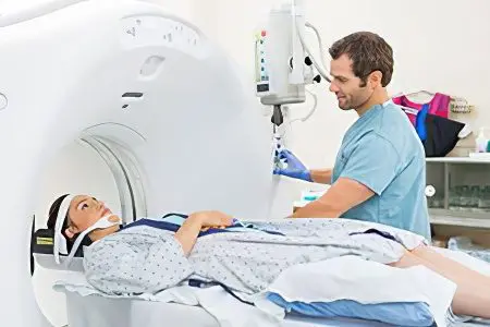
Identifying an aneurysm of the cerebral vessels can be quite problematic, since it often does not give any symptoms. Almost any specialist can suspect this pathology, which a sick person has to go through a lot. This is not surprising, because headaches can be caused by hypertension, intoxication of the body and many other disorders. Moreover, even such a common symptom as headache does not always occur in people with an aneurysm.
The doctor must definitely suspect the presence of any pathology of the central nervous system if the patient makes the following complaints or has symptoms such as:
Deterioration of visual, olfactory and / or auditory function;
The presence of seizures;
Loss of skin sensitivity;
Paralysis;
Disorders of coordination of actions;
The appearance of hallucinations;
Incorrect pronunciation of words or their spelling, etc.
Nevertheless, doctors are armed with a number of techniques that allow timely detection of cerebral aneurysm, but it is necessary to start the examination with an examination of the patient who applied for an appointment.
Examination of a patient with suspected aneurysm

Naturally, a routine examination will not allow identifying and diagnosing “cerebrovascular aneurysm”.
However, the doctor is able to suspect this pathology and send the patient for a more thorough examination:
Palpation allows you to assess the condition of the skin, as well as to suspect the presence of systemic diseases of the connective tissue. It is known that they often cause the formation of aneurysms.
With percussion, the doctor will not be able to detect an aneurysm, but this method makes it possible to detect other diseases that may accompany a defect in the cerebral vessels.
Listening to body noises allows you to detect pathological sounds that occur in the region of the heart, aorta, carotid artery. Taken together, these diagnostic criteria may prompt the doctor to think about the need for a thorough examination of the cerebral vessels.
Determination of blood pressure level. It is known that elevated blood pressure is a factor predisposing to the development of an aneurysm. In the case when the patient already knows his diagnosis, he must measure the pressure every day. Often, it is this manipulation that makes it possible to prevent or timely detect an aneurysm rupture.
Examination of the neurological status. During it, the doctor assesses the state of the patient’s reflexes (skin and muscle-tendon), tries to detect pathological reflexes. In parallel, the doctor evaluates the possibility of a person performing certain movements, the presence or absence of skin sensitivity. It is possible that the doctor will conduct an examination to detect meningeal symptoms.
The data obtained during the examination cannot serve as a basis for an accurate diagnosis. It is important to differentiate it from a brain tumor, from a transient ischemic attack, from an arteriovenous malformation, since all these pathological conditions give the same symptoms.
Tomography as a method for diagnosing aneurysms. CT and MRI can be called the leading methods for detecting this defect in cerebral vessels. However, they have some limitations. So, computed tomography is not prescribed for pregnant women, young children, patients with blood diseases and cancers. For a healthy adult, the dose of radiation that he receives during a CT scan is not dangerous.
As for MRI, this study is safe in terms of radiation, but it is not indicated for all patients. For example, it is not carried out in the presence of a metal-based implant or electronic prosthesis in the human body. Also, MRI is contraindicated in patients with a pacemaker.
After performing a computed or magnetic resonance imaging, the doctor will be able to obtain the following information about the aneurysm of the cerebral vessels, if any:
Its localization;
Its dimensions;
The presence of a thrombus;
Information about the number of aneurysms;
Information about the state of the brain tissues surrounding the aneurysm and about the speed of blood flow.
X-ray examination. Although the accuracy of angiography (X-ray examination with the introduction of a contrast agent into the vessels) is somewhat lower than CT and MRI, in most cases it allows visualization of the existing bulging of the vascular wall. The most informative angiography in the early development of the disease, which allows you to distinguish between a brain tumor and aneurysm of its vessels. However, CT and MRI are the most preferred methods for diagnosing this disease. Angiography is not recommended for pregnant women, children, patients with kidney disease.
EEG. Conducting an EEG does not allow you to make a diagnosis, but only provides information about the activity of certain parts of the brain. However, for an experienced doctor, it can be of value and make him think about the need for more complex diagnostic measures, such as MRI. In addition, EEG is absolutely safe for a person of any age and can be performed even for small children.
Treatment of cerebral aneurysm
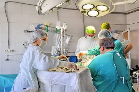
Surgery is the leading treatment for an aneurysm. It will remove the formation itself and restore the integrity of the vessels.
Surgery is the only effective treatment for cerebral aneurysms. If the size of the defect is more than 7 mm, then surgical treatment is mandatory. Emergency surgery is required for patients with a ruptured aneurysm. The following types of surgery are possible:
Direct microsurgical intervention
This type of surgery is also called aneurysm clipping. It is this that is most often implemented in the practice of microsurgery. The operation requires a craniotomy. The procedure itself lasts for many hours and carries high risks to the health and life of the patient.
Clipping steps:
trepanation of the skull;
Opening of the meninges;
Separation of the aneurysm from intact tissue;
Applying a clip on the body or neck of the aneurysm (this is necessary in order to remove it from the general blood flow);
Suturing.
To perform the operation, the doctor needs microsurgical equipment. In most cases, surgery is completed successfully, however, no doctor is able to guarantee a favorable prognosis.
In addition to clipping, a direct microsurgical wrapping operation can be performed, when the damaged vessel is strengthened using special gauze or a piece of muscle tissue for this purpose.
Endovascular operations
These surgeries are high-tech and do not require craniotomy. The aneurysm can be accessed with a needle that reaches the brain through the carotid or femoral artery and closes the existing lumen with a balloon or microcoil. They are fed through a needle through a catheter. As a result, the aneurysm is excluded from the general circulation. The whole procedure is carried out under the control of a tomograph.
Another type of endovascular surgery is aneurysm embolization using a special substance that hardens and prevents it from filling with blood. This procedure is carried out under the control of X-ray equipment with the introduction of a contrast agent.
If the hospital is equipped with equipment that allows for endovascular operations, then they should be given preference.
This is due to the following advantages of such methods:
Operations are low-traumatic;
Most often, the patient does not require the introduction of general anesthesia;
Craniotomy is not required;
The time spent by the patient in the hospital is reduced;
If the aneurysm is located in the deep tissues of the brain, then it will be possible to “neutralize” it only with the help of endovascular surgery.
combined operation.
This method involves a combination of the surgical method with endovascular technology. For example, a vessel can be occluded with a balloon followed by clipping; in general, there can be many options.
It should be understood that any operation is fraught with certain risks. This also applies to high-tech methods.
Among the most common complications are:
Spasms of blood vessels;
Rupture of the aneurysm with a balloon or coil;
Hypoxia;
Embolism of the vessel with blood clots;
Rupture of the aneurysm during the operation;
The death of the patient on the surgical table.
Video of the operation “endovascular embolization”, which uses natural access to the brain through the arteries to diagnose and treat a brain aneurysm:


