Contents

All living organisms on Earth are made up of cells, and each cell is surrounded by a protective shell – a membrane. However, the functions of the membrane are not limited to protecting organelles and separating one cell from another. The cell membrane is a complex mechanism that is directly involved in reproduction, regeneration, nutrition, respiration, and many other important cell functions.
The term “cell membrane” has been used for about a hundred years. The word “membrane” in translation from Latin means “film”. But in the case of a cell membrane, it would be more correct to speak of a combination of two films connected to each other in a certain way, moreover, different sides of these films have different properties.
Of decisive importance in this definition is not that the cell membrane separates one cell from another, but that it ensures its interaction with other cells and the environment. The membrane is a very active, constantly working structure of the cell, on which many functions are assigned by nature. From our article, you will learn everything about the composition, structure, properties and functions of the cell membrane, as well as the danger posed to human health by disturbances in the functioning of cell membranes.
History of cell membrane research
In 1925, two German scientists, Gorter and Grendel, were able to conduct a complex experiment on human red blood cells, erythrocytes. Using osmotic shock, the researchers obtained the so-called “shadows” – empty shells of red blood cells, then put them in one pile and measured the surface area. The next step was to calculate the amount of lipids in the cell membrane. With the help of acetone, the scientists isolated lipids from the “shadows” and determined that they were just enough for a double continuous layer.
However, during the experiment, two gross errors were made:
The use of acetone does not allow all lipids to be isolated from the membranes;
The surface area of the “shadows” was calculated by dry weight, which is also incorrect.
In 1935 year another pair of researchers, Danielly and Dawson, after long experiments on bilipid films, came to the conclusion that proteins are present in cell membranes. There was no other way to explain why these films have such a high surface tension. Scientists have presented to the public a schematic model of a cell membrane, similar to a sandwich, where the role of slices of bread is played by homogeneous lipid-protein layers, and between them instead of oil is emptiness.
In 1950 year With the help of the first electron microscope, the Danielly-Dawson theory was partially confirmed – microphotographs of the cell membrane clearly showed two layers consisting of lipid and protein heads, and between them a transparent space filled only with tails of lipids and proteins.
In 1960 yearGuided by these data, the American microbiologist J. Robertson developed a theory about the three-layer structure of cell membranes, which for a long time was considered the only true one. However, as science developed, more and more doubts were born about the homogeneity of these layers. From the point of view of thermodynamics, such a structure is extremely unfavorable – it would be very difficult for cells to transport substances in and out through the entire “sandwich”. In addition, it has been proven that the cell membranes of different tissues have different thickness and method of attachment, which is due to different functions of organs.
In 1972 year microbiologists S.D. Singer and G.L. Nicholson was able to explain all the inconsistencies of Robertson’s theory with the help of a new, fluid-mosaic model of the cell membrane. Scientists have found that the membrane is heterogeneous, asymmetric, filled with fluid, and its cells are in constant motion. And the proteins that make up it have a different structure and purpose, in addition, they are located differently relative to the bilipid layer of the membrane.
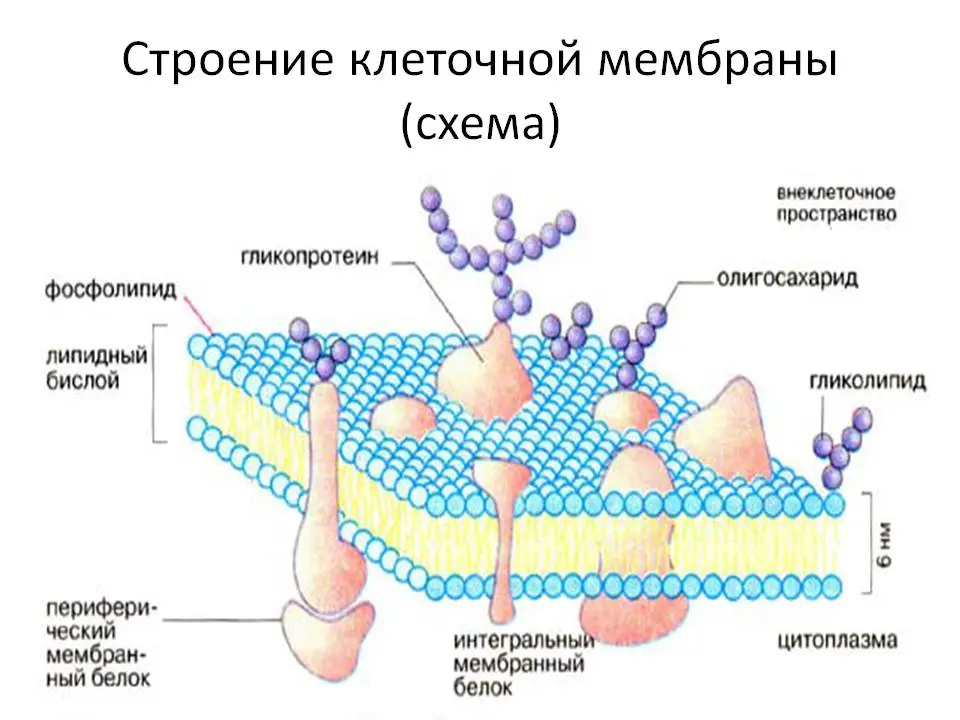
Cell membranes contain three types of proteins:
Peripheral – are attached to the surface of the film;
semi-integral – partially penetrate the bilipid layer;
Integral – completely penetrate the membrane.
Peripheral proteins are associated with the heads of membrane lipids through electrostatic interaction, and they never form a continuous layer, as was previously believed. And semi-integral and integral proteins serve to transport oxygen and nutrients into the cell, as well as to remove decay products from it and more for several important features, which you will learn about later.
Properties and functions of the cell membrane

The cell membrane performs the following functions:
barrier – the permeability of the membrane for different types of molecules is not the same. To bypass the cell membrane, the molecule must have a certain size, chemical properties and electric charge. Harmful or inappropriate molecules, due to the barrier function of the cell membrane, simply cannot enter the cell. For example, with the help of the peroxide reaction, the membrane protects the cytoplasm from peroxides that are dangerous for it;
Transport Passive, active, regulated and selective exchange passes through the membrane. Passive metabolism is suitable for fat-soluble substances and gases consisting of very small molecules. Such substances penetrate into and out of the cell without energy expenditure, freely, by diffusion. The active transport function of the cell membrane is activated when necessary, but difficult to transport substances need to be carried into or out of the cell. For example, those with a large molecular size, or unable to cross the bilipid layer due to hydrophobicity. Then protein pumps begin to work, including ATPase, which is responsible for the absorption of potassium ions into the cell and the ejection of sodium ions from it. Regulated transport is essential for secretion and fermentation functions, such as when cells produce and secrete hormones or gastric juice. All these substances leave the cells through special channels and in a given volume. And the selective transport function is associated with the very integral proteins that penetrate the membrane and serve as a channel for the entry and exit of strictly defined types of molecules;
matrix – the cell membrane determines and fixes the location of organelles relative to each other (nucleus, mitochondria, chloroplasts) and regulates the interaction between them;
Mechanical – ensures the restriction of one cell from another, and, at the same time, the correct connection of cells into a homogeneous tissue and the resistance of organs to deformation;
Protective – both in plants and animals, the cell membrane serves as the basis for building a protective frame. An example is hard wood, dense peel, prickly thorns. In the animal world, there are also many examples of the protective function of cell membranes – turtle shell, chitinous shell, hooves and horns;
Energy — the processes of photosynthesis and cellular respiration would be impossible without the participation of cell membrane proteins, because it is with the help of protein channels that cells exchange energy;
receptor— proteins embedded in the cell membrane may have another important function. They serve as receptors through which the cell receives a signal from hormones and neurotransmitters. And this, in turn, is necessary for the conduction of nerve impulses and the normal course of hormonal processes;
Enzymatic – Another important function inherent in some proteins of cell membranes. For example, in the intestinal epithelium, digestive enzymes are synthesized with the help of such proteins;
Biopotential – the concentration of potassium ions inside the cell is much higher than outside, and the concentration of sodium ions, on the contrary, is greater outside than inside. This explains the potential difference: inside the cell the charge is negative, outside it is positive, which contributes to the movement of substances into the cell and out in any of the three types of metabolism – phagocytosis, pinocytosis and exocytosis;
marking – on the surface of cell membranes there are so-called “labels” – antigens consisting of glycoproteins (proteins with branched oligosaccharide side chains attached to them). Since side chains can have a huge variety of configurations, each type of cell receives its own unique label that allows other cells in the body to recognize them “by sight” and respond to them correctly. That is why, for example, human immune cells, macrophages, easily recognize a foreigner that has entered the body (infection, virus) and try to destroy it. The same thing happens with diseased, mutated and old cells – the label on their cell membrane changes and the body gets rid of them.
Cellular exchange occurs across membranes, and can be carried out using three main types of reactions:
Phagocytosis – a cellular process in which phagocyte cells embedded in the membrane capture and digest solid particles of nutrients. In the human body, phagocytosis is carried out by membranes of two types of cells: granulocytes (granular leukocytes) and macrophages (immune killer cells);
Pinocytosis – the process of capture by the surface of the cell membrane of the molecules of the liquid in contact with it. For nutrition by the type of pinocytosis, the cell grows thin fluffy outgrowths in the form of antennae on its membrane, which, as it were, surround a drop of liquid, and a bubble is obtained. First, this vesicle protrudes above the surface of the membrane, and then it is “swallowed” – it hides inside the cell, and its walls merge with the inner surface of the cell membrane. Pinocytosis occurs in almost all living cells;
Exocytosis – the reverse process, in which vesicles are formed inside the cell with a secretory functional fluid (enzyme, hormone), and it must somehow be removed from the cell into the environment. To do this, the vesicle first merges with the inner surface of the cell membrane, then bulges outward, bursts, expels the contents and again merges with the surface of the membrane, this time from the outside. Exocytosis takes place, for example, in the cells of the intestinal epithelium and the adrenal cortex.
The structure of the cell membrane
Cell membranes contain three classes of lipids:
Phospholipids;
Glycolipids;
Cholesterol.

Phospholipids (a combination of fats and phosphorus) and glycolipids (a combination of fats and carbohydrates), in turn, consist of a hydrophilic head, from which two long hydrophobic tails extend. But cholesterol sometimes occupies the space between these two tails and does not allow them to bend, which makes the membranes of some cells rigid. In addition, cholesterol molecules streamline the structure of cell membranes and prevent the transition of polar molecules from one cell to another.
But the most important component, as can be seen from the previous section on the functions of cell membranes, are proteins. Their composition, purpose and location are very diverse, but there is something in common that unites them all: annular lipids are always located around the proteins of cell membranes. These are special fats that are clearly structured, stable, have more saturated fatty acids in their composition, and are released from membranes along with “sponsored” proteins. This is a kind of personal protective shell for proteins, without which they simply would not work.
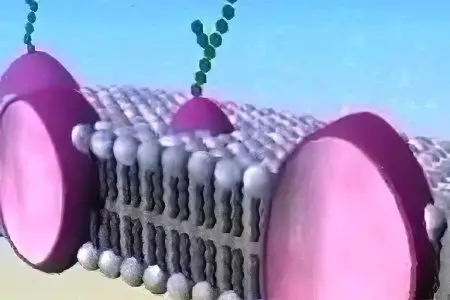
The structure of the cell membrane is three-layered. A relatively homogeneous liquid bilipid layer lies in the middle, and proteins cover it on both sides with a kind of mosaic, partially penetrating into the thickness. That is, it would be wrong to think that the outer protein layers of cell membranes are continuous. Proteins, in addition to their complex functions, are needed in the membrane in order to pass inside the cells and transport out of them those substances that are unable to penetrate the fat layer. For example, potassium and sodium ions. For them, special protein structures are provided – ion channels, which we will discuss in more detail below.
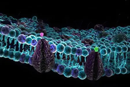
If you look at the cell membrane through a microscope, you can see a layer of lipids formed by the smallest spherical molecules, along which, like the sea, large protein cells of various shapes float. Exactly the same membranes divide the internal space of each cell into compartments in which the nucleus, chloroplasts and mitochondria are comfortably located. If there were no separate “rooms” inside the cell, the organelles would stick together and would not be able to perform their functions correctly.
As can be seen from this definition, the membrane is the most important functional component of any cell. Its significance is as great as that of the nucleus, mitochondria and other cell organelles. And the unique properties of the membrane are due to its structure: it consists of two films stuck together in a special way. Molecules of phospholipids in the membrane are located with hydrophilic heads outward, and hydrophobic tails inward. Therefore, one side of the film is wetted by water, while the other is not. So, these films are connected to each other with non-wettable sides inward, forming a bilipid layer surrounded by protein molecules. This is the very “sandwich” structure of the cell membrane.
Ion channels of cell membranes
Let us consider in more detail the principle of operation of ion channels. What are they needed for? The fact is that only fat-soluble substances can freely penetrate through the lipid membrane – these are gases, alcohols and fats themselves. So, for example, in red blood cells there is a constant exchange of oxygen and carbon dioxide, and for this our body does not have to resort to any additional tricks. But what about when it becomes necessary to transport aqueous solutions, such as sodium and potassium salts, through the cell membrane?
It would be impossible to pave the way for such substances in the bilipid layer, since the holes would immediately tighten and stick together back, such is the structure of any adipose tissue. But nature, as always, found a way out of the situation and created special protein transport structures.
There are two types of conductive proteins:
Transporters – semi-integral protein pumps;
Channel formers are integral proteins.
Proteins of the first type are partially immersed in the bilipid layer of the cell membrane, and look out with their heads, and in the presence of the desired substance, they begin to behave like a pump: they attract the molecule and suck it into the cell. And proteins of the second type, integral, have an elongated shape and are located perpendicular to the bilipid layer of the cell membrane, penetrating it through and through. Through them, as through tunnels, substances that are unable to pass through fat move into and out of the cell. It is through ion channels that potassium ions penetrate into the cell and accumulate in it, while sodium ions, on the contrary, are brought out. There is a difference in electrical potentials, so necessary for the proper functioning of all the cells of our body.
[Tutorial video] The structure of the cell’s plasma membrane:


