Contents
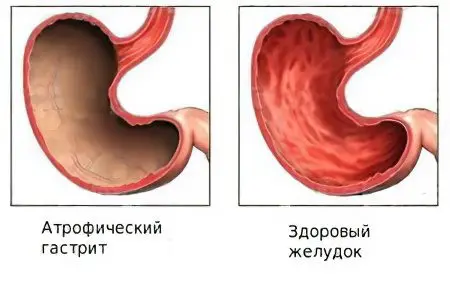
Atrophic gastritis, the most insidious type of chronic gastritis, is a likely cause of a precancerous condition of the stomach. It develops more often in middle-aged and older men. At the onset, inflammation is asymptomatic. With the depletion of compensatory mechanisms, it does not always have a vivid clinical picture.
What is atrophic gastritis?
The absence of vivid symptoms at the first stage of pathogenesis is not a favorable sign. On the contrary, a person who does not experience obvious discomfort does not attach importance to the problem. In vain. Let’s try to explain in a simple and accessible way the insidiousness of this disease.
The key word in the name of the disease is atrophy. This means that the cells of the walls of the stomach, which are part of the secretory glands, undergo atrophic degeneration during the course of the disease, that is, they lose the ability to function normally and do not produce components of gastric juice. It has been proven that, first of all, the glands are transformed into simpler formations that produce mucus instead of gastric juice. Usually atrophic gastritis occurs against the background of low acidity of the stomach.
However, the main danger of atrophic gastritis is not associated with a change in the acidity of gastric juice, since the pH level can be corrected. The danger is elsewhere. Atrophic gastritis has a status generally recognized by the medical community as a provocateur of stomach cancer in humans.
So, in order. All cells of the body, including the cells of the walls of the stomach, are in every second cooperation with the body. This means that regeneration – origin, morphological and functional differentiation, functional load, natural cell death and their subsequent renewal are influenced by hormonal, immune, enzymatic and other, yet unknown to science, regulatory factors. Until now, no one has been able to reliably and radically change the properties of mature body cells. Normally, all cells of the organs of the body have a strict specialization – this is an axiom of modern biological science.
The pathogenesis of atrophic gastritis

To simplify the task, we describe pathogenesis as a two-stage process. We agree that at the first stage of pathogenesis acid-resistant bacteria play a leading role, and at the second stage autoimmune processes of the body play a leading role.
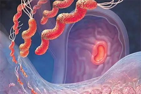
In many forms of gastritis, the cells of the glands of the inner walls of the stomach are attacked by the bacteria Helicobacter pylori, which damage them and locally change the pH of the environment of the walls of the stomach. Bacteria are common inhabitants of the acidic environment of the stomach. They only create the soil, open the gate for the development of gastritis by atrophic and any other type of inflammation.
At the second stage of atrophic gastritis, complex autoimmune processes are involved in the pathogenesis, which affect the immature forms of gland cells and suppress their subsequent specialization. The mechanism of the origin and course of autoimmune reactions is of interest to scientists, but in this text their disclosure is not of fundamental importance.
Suppression of cell specialization is the key word in the pathogenesis of this type of inflammation. This means that the cells of the glands of the walls of the stomach under the influence of autoimmune reactions atrophy, cease to perform the complex work of producing the components of gastric juice.
The physiological process of regeneration of the glandular cells of the stomach is disrupted. Regeneration means that normally the place of glandular cells that have exhausted their vital resource is occupied by new cells with similar properties. In a healthy body, a complete renewal of the cells of the mucous membranes of the stomach occurs every six days.
As a result of impaired regeneration, glandular cells instead of hydrochloric acid begin to produce a simpler product – mucus. This mucus has protective properties, but is poorly involved in digestion. Therefore, the walls of the stomach, abundantly covered with mucus, have the appearance of healthy tissue during a routine endoscopic examination. The environment of the stomach from acidic is transformed into slightly acidic, up to achilia.
Subsequently, under the influence of an autoimmune cascade of reactions, damaged cells begin to produce a large number of immature cells similar to themselves, which are unable to develop and have finally lost the ability to acquire secretory specialization. In this case, it is pathological regeneration. Conventionally, such immature cells can be called the now fashionable term – stem cells.
Any healthy person has stem cells, but in a normally functioning organism they invariably acquire properties strictly specified by evolutionary memory and are transformed into mature cells: stomach, intestines, heart, lungs, other organs and tissues, and perform exclusively specific functions for each cell type.
If scientists learn how to control stem cells with certainty, this will mean a revolution, and will allow humanity to embark on the path of individually regulated lifespan. It will be possible to grow any organ or tissue, and thereby change metabolic processes, hormonal levels, and so on. So far, work on stem cell management is at the initial stage of scientific study, and the practical application of this technique is a guaranteed risk. But back to the topic of atrophic gastritis.
It is believed that atrophy of the cells of the gastric walls cannot be completely cured. However, the correct drug exposure, adherence to diet, exclusion from the diet of certain types of food significantly reduce the risk of oncological processes. Regarding the diagnosis, prevention of atrophic gastritis and the possible risk of developing oncological processes, you should consult with your doctor.
In a fatal combination of circumstances, that is, a strong, external and / or internal impact, an explosive, growing exponentially, growth of young (stem) cells of the walls of the stomach is provoked.
These cells do not carry a functional load that is useful for the body, on the contrary, they destroy it. The only function of imperfect cells that do not have a cooperative relationship with the body is the constant, unregulated by the body reproduction of their own similar pathological (cancerous) cells and a negative impact on the body through metabolic products.
It should be recalled that the pathogenesis described above is a simplified representation of the true pathogenesis of atrophic gastritis. The text does not mention serious morphological damage to the gastric glands, changes in hormonal, vitamin and other types of metabolism, the influence of autoimmune processes on the development of pathogenesis and the influence of dystrophic processes on pathogenesis. There is no mention of the greater or lesser influence of certain strains of acid-fast bacteria and duodenogastric reflux on chronic gastritis. In a schematic, generalized form, an idea is given of the transformation of atrophic gastritis into a precancerous condition.
Symptoms of atrophic gastritis

The vast majority of serious researchers indicate the absence of any significant symptoms of atrophic gastritis at the first stage of pathogenesis. Many have noticed the absence of a bright pain syndrome in atrophic gastritis, which is characteristic of hyperacid gastritis. Pain is absent at all stages of atrophic gastritis.
Often mentioned symptoms at the stage of depletion of the body’s compensatory mechanisms include signs that are common to all types of gastritis. During a clinical examination, patients complain of a feeling of heaviness in the solar plexus after eating, regardless of its volume.
There are also complaints about the following signs of gastrointestinal pathology:
belching;
nausea;
heavy odor from the mouth;
flatulence;
overflows, rumbling;
constipation is recorded more often than diarrhea;
Symptoms that are not directly related to disorders of the gastrointestinal tract include:
decreased body weight;
hypovitaminosis (a clear decrease in the level of cyanocobalamin (vitamin B12), manifests itself in the form of anemia, ulcerations on the mucous membrane of the mouth, tingling of the tongue, headaches, jaundice of the skin);
hormonal metabolism disorders (hypocorticism, decreased libido)
However, the main signs of atrophic gastritis are detected in laboratory, functional and instrumental studies.
It should be said that ultrasound, radiography, CT of the abdominal cavity without contrast agents, MRI do not provide comprehensive information about the pathology. The greatest diagnostic value is the methods of endoscopy, gastroscopy and its varieties, for example, chromogastroscopy. This is a method for examining the walls of the stomach after preliminary coloring of their surface.
With the help of a gastroscope, thinning and smoothing of the walls are observed. The vessels of the gastric walls are clearly visible (normally they are not visible). Wall biopsy studies reveal dystrophy and atrophy of the gastric glands. Valuable is the method of intragastric pH measurement. Almost always, a change in the pH of the stomach environment towards a neutral reaction, up to achilia, is detected. The list of mandatory methods for diagnosing atrophic gastritis includes the study of the microflora of the stomach. Many experts consider the routine detection of Helicobacter pylori bacteria to be an uninformative diagnostic method.
The most convenient, promising, non-invasive (sparing) method of blood testing for the state of the functional activity of the stomach is a gastropanel.
Gastropanel is a blood test method that is based on the identification of:
Antibodies to Helicobacter pylori;
Pepsinogen I – the protein responsible for the production of HCL;
Gastrin 17 – a hormone that regulates the secretion of hydrochloric acid, regeneration and motility of the walls.
It is believed that the gastropanel should be used in combination with histological studies of the cells of the stomach wall. Comparison of their results provides very valuable diagnostic information.
Types of atrophic gastritis

In-depth laboratory, instrumental and other studies are valuable in determining the types of atrophic gastritis, depending on the location of the pathogenesis and the nature of the damage. Studies are valuable for identifying and differentiating various pathological formations in the stomach, stages and forms of its inflammation.
Acute atrophic gastritis
In this case, we should talk about the stage of exacerbation of chronic atrophic inflammation of the walls of the stomach. In some sources, this condition is called active gastritis. Symptoms resemble manifestations of acute superficial inflammation of the stomach.
Laboratory and instrumental methods establish the following characteristic signs of acute atrophic gastritis:
swelling of the walls of the body;
plethora of vessels of the walls;
infiltration of leukocytes outside the blood vessels;
destruction of the integumentary epithelium, rarely – erosion on the mucous membrane.
In some cases, atrophy of glandular tissue cells occurs under the influence of external emergency factors – strong acids, alkalis, chemical poisons, and so on. Diagnosis and therapy of acute toxic atrophy of the glandular tissue of the stomach is carried out not by gastroenterologists, but by doctors specializing in toxicology, narcology and surgery.
Symptoms of acute atrophic gastritis are varied: severe pain, vomiting, diarrhea, fever, impaired consciousness – fainting, coma. Other specific symptoms are characteristic of each specific pathological process. The impact on the mucous membranes of strong pathogens often ends in the death of the patient due to general intoxication of the body, cardiac or respiratory arrest.
Chronic atrophic gastritis
It is an independent disease, and not a transformation of acute gastritis. This condition is sometimes referred to as inactive gastritis or gastritis in remission. It is characterized by a long-term, progressive atrophy of glandular tissue cells, the predominance of dystrophic processes over inflammatory ones. Pathogenesis leads to changes in secretory, motor and absorption functions. In the chronic form of atrophic gastritis, anatomically associated with the stomach organs are involved in the pathogenesis: the duodenum, esophagus, as well as organs associated with the stomach functionally: liver, pancreas, endocrine glands. Due to the general intoxication of the body, the process of hematopoiesis and the nervous system are involved in the pathogenesis.
Pathogenesis, as a rule, develops against the background of low acidity of gastric juice. Clinical symptoms correspond to gastritis with low acidity.
The diagnosis of acute and chronic gastritis is made on the basis of differential diagnosis data. The examination is carried out using instrumental, functional and laboratory methods. Of particular value are endoscopy and its varieties, pH-metry, histological methods for examining a biopsy specimen, laboratory blood tests – a gastropanel.
In the course of diagnostic studies, chronic atrophic gastritis is manifested by the following symptoms:
normal or thinned organ wall;
smoothed mucous membrane;
wide gastric dimples;
flattening of the epithelium;
low secretory activity of the glands;
moderate infiltration of leukocytes outside the vessels;
degeneration (vacuolization) of gland cells.
Focal atrophic gastritis

It is characterized by the appearance of foci of pathologically altered tissue of the walls of the stomach. Acute focal gastritis in some cases occurs against the background of increased acidity of gastric juice. Probably, areas of glandular tissue not involved in the pathogenesis compensate for the functions of damaged foci by increasing the secretion of hydrochloric acid. Otherwise, the symptoms of the disease practically do not differ from the symptoms of ordinary gastritis.
In the subclinical course, focal atrophic gastritis is manifested by intolerance to certain products: usually these are dishes based on milk, fatty meat, eggs. After their use, heartburn, nausea, and sometimes vomiting begin. Differential diagnosis is made on the basis of laboratory and instrumental studies.
Moderate atrophic gastritis
According to the degree of involvement of glandular tissue in degenerative-atrophic processes, a moderate form of inflammation is sometimes isolated in clinical practice. The designation is conditional and implies a mild, partial form of pathological transformation of the cells of the gastric walls.
Moderate atrophic gastritis is detected only by histological examination of glandular cells. At the same time, the number of undamaged cells per unit area of the gastric mucosa is determined, and the depth of microstructural changes in the glandular and degenerate tissue is analyzed, which serves as a criterion for determining this type of disease.
Clinical symptoms correspond to the usual dyspeptic disorders. The pain characteristic of acute forms of gastritis is not always fully manifested in this disease. More often, patients complain of a feeling of discomfort in the epigastrium that occurs after eating. Pain is possible only when eating heavy (spicy, salty, smoked, pickled or fatty) food.
Superficial atrophic gastritis
In accordance with the working classification – a harbinger of atrophic inflammation of the stomach. This is an early stage of chronic inflammation. Damage is minimal, clinical symptoms are not expressed. Differential diagnosis is possible only with the help of endoscopy. A detailed study establishes:
normal thickness of the stomach wall;
moderate degeneration of the integumentary epithelium;
slight hypersecretion of cells.
Antral atrophic gastritis
The antrum is located in the lower part of the stomach, closer to the exit from the organ, and is adjacent to the duodenum. The disease is characterized by scarring of the antrum. Visually, this section looks like a tube with compacted walls. Condensation and tension are called rigidity. This form of gastritis is characterized by moderately pronounced clinical signs of dyspepsia – dull pain in the solar plexus, as well as:
nausea in the morning;
belching after eating;
loss of appetite;
weight loss;
general weakness.
When measuring the pH level, its normal value is rarely set – more often a decrease in the slightly acidic direction. An instrumental study of the mucous membranes reveals deformation, pronounced macroscopic changes on the inner walls of the organ, and a decrease in the peristalsis of the walls due to their rigidity. Macroscopic changes are often diagnosed as tumor formations on the mucous membrane. Ulcerative processes are often diagnosed in the antrum of the stomach.
Diffuse atrophic gastritis
Means the absence of serious dystrophic changes. This form of inflammation is an intermediate link, a transitional stage between superficial and dystrophic damage to the walls. The main sign of diffuse gastritis is the presence of local foci of degeneration of the glands of the walls of the stomach, as well as immature cells with signs of impaired secretory activity.
Other signs of diffuse atrophic gastritis:
rollers on the walls of the stomach;
deepened gastric pits;
microstructural damage to gland cells.
Treatment of atrophic gastritis
Due to the variety of microstructural manifestations of atrophic gastritis and the sparse clinical symptoms, there is no single approach to the treatment of this disease. It is recognized that the formed atrophic process cannot be corrected. That is, degenerated cells cannot be transformed back into glandular ones.
Meanwhile, effective schemes of drug treatment of atrophic gastritis in various forms and at different stages have been proposed and exist, preventing the further development of pathogenesis.
All treatment schemes are based on the results of an in-depth study of the body. This is very important, as different data suggest different therapeutic approaches. In this article, we will not concretize treatment methods. Let the attending physician do this, based on specific conditions, the state of the patient’s body, and the involvement of various links in the chain in the pathogenesis.
Meanwhile, the traditional treatment regimen for atrophic gastritis includes:
Eradication of Helicobacter pylori if acid-fast bacteria have a marked effect on pathogenesis. Helicobacter pylori eradication methods are constantly being improved.
Eradication tasks:
suppression of the development of bacteria and prevention of the formation of their resistance to antibiotics;
the use of proton pump inhibitors to improve well-being;
reduction in the duration of treatment;
reduction in the number of drugs, which significantly reduces the number of side effects from treatment;
Usually, three- and four-component eradication schemes are used:
As a means of suppressing the activity of bacteria, antibiotics (tetracycline, penicillin), as well as the antibacterial drug metronidazole (Trichopolum) are used. The dosage and frequency rate is indicated by the doctor.
Omeprazole, Lansoprazole, Esomeprazole, Rabeprazole, Pantoprazole, Ranitidine, bismuth citrate and others are used as proton pump inhibitors.
To influence the development of autoimmune processes in atrophic gastritis has not yet been fully learned. The use of hormonal drugs and other immunocorrectors in most cases is not justified.
Pathogenetic therapy of atrophic gastritis involves the complex use of drugs from various groups, among them:
means that facilitate gastric digestion – preparations of hydrochloric acid and enzymes of gastric juice.
in conditions of insufficiency of vitamins of group B12 use appropriate vitamin preparations in the form of parenteral injections.
means that affect the production of hydrochloric acid in the form of mineral waters (Essentuki 4,17 and others). Although they are not drugs, they show high therapeutic activity in some cases.
drugs that reduce inflammation – psyllium juice or granular pharmacological preparation from psyllium (Plantaglucid).
In recent years, more often began to be used in the treatment of gastrointestinal inflammation Riboxin. This drug has properties useful in the treatment of atrophic gastritis.
to protect the mucous membrane, bismuth or aluminum preparations are used (Bismuth nitrate basic, Vikalin, Vikair or Rother, Kaolin).
drugs that regulate the motor function of the stomach. Among the drugs of this pharmacological group, Domperidone and Cisapride are most often used.
All of the above drugs are prescribed during the active phase of inflammation of the stomach with symptoms of atrophy. During the period of remission, the main principle of treatment is to replenish the missing substances for proper digestion.
Diet for atrophic gastritis

Dietary nutrition is an integral part of the treatment of all types of gastritis. Treatment of atrophic gastritis (AG) is associated with some difficulties in the organization of nutrition. Depending on the objectives of therapy, four types of diets are recommended, developed by nutritionist M.I. Pevzner.
The basic diet for atrophic gastritis is diet number 2. It involves the full nutrition of the patient and the stimulation of functional glands. Recommended dishes must be boiled, easily fried, stewed, baked. Do not use chilled products with a rough structure. The diet allows the use of a variety of dishes: meat, fish. Sour-milk, flour products, hard-boiled eggs and in the form of an omelet are allowed. Vegetables and fruits are widely used. In total, more than thirty types of different products are allowed, allowing you to organize a high-quality and varied diet.
With severe pain, a different diet is prescribed. It is designated No. 1a and is prescribed in the first days of the disease. This diet option provides a minimal burden on digestion. The task of the diet is to reduce the reflex excitability of the gastric mucosa. Foods that have a stimulating effect on the receptors of the stomach are excluded from the diet. Food is allowed only in the form of a liquid or mashed potatoes, steamed, boiled, mashed. The diet consists of nine main recommended dishes, mostly mashed soups. Dairy products are also allowed, provided they are well tolerated – whole milk, cream, cottage cheese.
Diet number 1 is prescribed after the symptoms of inflammation subside. It is used to speed up the recovery process of the inflamed gastric mucosa. This diet contributes to the normalization of the secretory and motor functions of the stomach. Hot and cold dishes are excluded from the menu. Fiber-rich foods are not recommended. The list of diet includes about eleven items of dishes.
Diet number 4 is prescribed for severe enteral syndromewhen there is an individual intolerance to milk and other products. The task of this diet is to normalize the functioning of the stomach by reducing inflammation in the gastric mucosa. Fractional diet. After the inflammation subsides, they always return to good nutrition. With atrophic gastritis, this is the number two diet.


