Contents
Angiopathy of the vessels of the retina – what is it?
The retina of the eye needs a large intake of nutrients and oxygen, as it is responsible for capturing light waves, converting them into a nerve impulse and transmitting them to the brain, where image formation takes place. Lack of blood supply to the choroid causes serious visual impairment. Angiopathy of the retinal vessels is not a separate disease, but a pathology that develops as a result of the destruction of blood vessel cells and disruption of their functions in diseases of various genesis.
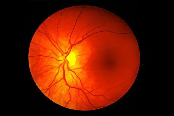
Angiopathy of the retinal vessels – this is a pathological violation of the tone of the blood vessels and capillaries of the fundus. As a result, their tortuosity, narrowing or expansion occurs. There is a change in the speed of blood flow and a failure of nervous regulation. Vascular defects make it possible to suspect and diagnose the underlying disease before its clinical manifestations.
Pathology of this type signals the presence in the body of a disease that prevents normal blood circulation, affects the tone of small and large vessels, causes necrotic lesions of a certain area of the retina, threatens with complete or partial loss of vision or a decrease in its quality. Angiopathy is more common in adult patients (over 35 years old) against the background of chronic diseases, but is sometimes diagnosed in childhood, and even in newborns.
Causes of retinal angiopathy

The most important structure of the eye – the retina – quickly responds to the slightest disturbances in the blood supply system. Angiopathy is not an independent disease, it serves as a signal of a disease in which there is a negative effect on the eye vessels. Pathological processes in the body cause damage to the walls of the eye vessels, their modification and disruption of the structure.
The main reasons that lead to the occurrence of angiopathy:
Hypertonic disease. High blood pressure has a detrimental effect on the walls of the eye vessels, destroying their inner layer. The vascular wall thickens, and fibrotization occurs. There is a violation of blood circulation, the formation of blood clots and hemorrhages. Due to the constantly elevated pressure, some vessels burst. A characteristic sign of hypertensive angiopathy is convoluted, narrowed vessels of the fundus. In the first degree of hypertension, changes in the vessels of the eyes are observed in a third of patients, in the second degree – in half of the patients, and in the third stage of hypertension, the fundus vessels are modified in all patients;
Diabetes. The disease causes damage to the vascular walls not only in the retina, but throughout the body. Pathology develops against the background of constantly elevated blood glucose levels. This causes the development of occlusions, blood seepage into the retinal tissue, thickening and growth of the capillary wall, a decrease in the diameter of blood vessels and deterioration of blood microcirculation in the eyes. Pathogenesis often entails a gradual loss of vision;
Injuries to the skull, eyes and spine (cervical region), strong and prolonged compression of the chest. The condition leads to a sharp increase to high numbers of intracranial pressure, to rupture of the walls of blood vessels and hemorrhage into the retina;
Hypotension. A decrease in vascular tone entails branching of the vessels, their strong expansion, palpable pulsation, a decrease in blood flow velocity, and also contributes to the formation of blood clots in the retinal vessels, increases the permeability of the vessel walls.
Factors that contribute to the occurrence of dangerous angiopathy:
Increased intracranial pressure;
Bad habits (smoking, alcohol);
Poisoning (acute or chronic);
advanced age;
Congenital anomalies of the walls of blood vessels;
Osteochondrosis.
There are several more varieties of this pathology, which also sometimes occur:
Juvenile angiopathy. The inflammatory process in the vessels of the retina develops for an unknown reason. It is accompanied by small hemorrhages in the vitreous body of the eye and retina. The most severe type of the disease, which contributes to retinal detachment, also provokes the occurrence of cataracts and glaucoma, often leading to blindness;
Angiopathy in premature newborns. The disease is rare, the cause of its occurrence is a complication of childbirth or birth trauma. Retinal damage is characterized by proliferative changes in blood vessels, their narrowing and impaired blood flow;
Angiopathy during pregnancy. In the early stages, the disease does not carry threatening consequences, but in its advanced form it threatens with irreversible complications (retinal detachment). This pathology can develop in the second half of pregnancy against the background of hypertension or other diseases that are characterized by weakness of the vascular walls.
Any pathology or disease that negatively (directly or indirectly) affects the state of the vessels can lead to angiopathy.
The most common causes of angiopathy include:
Arterial hypertension of various etiologies;
Atherosclerosis;
Congenital pathologies of the vascular walls;
Systemic vasculitis;
Diabetes;
Increased intracranial pressure;
Traumatic lesions of the eyes;
Head bruises;
Some blood diseases;
Osteochondrosis of the cervical spine;
Intoxication of the body.
Additional risk factors:
Old age and presbyopia (senile vision);
Work in hazardous production;
Smoking and alcohol abuse;
Exposure to radiation.
Symptoms of retinal angiopathy

Vascular angiopathy is divided into types depending on the underlying disease:
Diabetic angiopathy. The most common. In patients with type 1 diabetes, it is observed in 40% of cases, in patients with type 2 – in 20%. Usually angiopathy begins to develop after 7-10 years from the onset of the disease. Two variants of development are possible: microangiopathy and macroangiopathy. With microangiopathy, capillaries are affected and thinner, which leads to impaired microcirculation and hemorrhages. With macroangiopathy, larger vessels are affected, occlusions (blockage) occur, which leads to retinal hypoxia;
Hypertensive angiopathy. Against the background of chronically elevated pressure, narrowing of the arteries of the retina and dilation of the veins occur. Vascular sclerosis is gradually formed, the venous bed becomes branched, exudates are formed due to blood seepage through the capillary walls;
Hypotonic angiopathy. Against the background of arterial hypotension, on the contrary, the arteries expand, the blood flow slows down, there is a pulsation of the veins, the vessels become tortuous, which increases the likelihood of blood clots. The characteristic symptoms in this case are a sensation of throbbing in the eyes and dizziness;
Traumatic angiopathy. With injuries to the head or chest, squeezing the abdomen, osteochondrosis, intraocular pressure can rise sharply. If the vessels do not withstand the load, then their ruptures occur with subsequent hemorrhages;
Angiopathy during pregnancy. In this case, angiopathy is functional in nature and disappears on its own after 2-3 months after childbirth. This is explained by the fact that an increase in the volume of circulating blood causes passive expansion of the retinal vessels. Another question is if diabetic or hypertensive angiopathy was present before pregnancy. In this case, it is likely to begin to progress rapidly.
The danger of angiopathy is that in the early stages and for quite a long time it is asymptomatic. At the stage of a noticeable deterioration in vision, the process is usually already irreversible.
General symptoms of angiopathy:
Decreased visual acuity;
The appearance of fog and spots before the eyes;
Narrowing of the field of view;
Feeling of pulsation in the eyeball;
The presence of burst vessels and yellow spots on the conjunctiva.
Additional symptoms:
Bleeding from the nose;
Pain in the legs;
Blood in the urine.
Treatment of retinal angiopathy

Treatment of angiopathy is carried out strictly individually for each patient, taking into account the nature of the disease and the severity. Drug therapy is aimed at the complete elimination of the factors that provoke this pathology: in the case of hypertension, antihypertensive drugs are prescribed, in diabetes mellitus – drugs that help lower blood sugar levels. Treatment of angiopathy of the retinal vessels is carried out in a complex by conservative and surgical methods with the interaction of many doctors: an ophthalmologist, an ophthalmologist, an internist, an endocrinologist, a cardiologist, a rheumatologist, and a neuropathologist.
Therapy of the pathological process includes conservative measures:
Taking drugs that improve blood microcirculation and strengthen the vascular walls: Trental, Pentoxifylline, Actovegin, Vasonite, Solcoseryl, Arbiflex, Cavinton;
The appointment of medications that reduce vascular permeability: Dobesilat, Parmidin, ginkgo biloba extract;
Vitamin therapy with drugs of group B (B6, B1, B12, B15), C, P, E;
Taking drugs that prevent thrombosis: Trombonet, Lospirin, Dipyridamole, Magnikor, Ticlodipine;
Drops to improve blood microcirculation in the eyes: Emoksipin, Taufon;
Medications for the treatment of a primary disease that provoked angiopathy of the vessels of the retina of the eye (hypotensive, hypoglycemic);
Physiotherapeutic procedures: laser irradiation, magnetic resonance therapy, acupuncture;
Traditional medicine recommends taking infusions of herbs: chamomile, St. John’s wort, yarrow, lemon balm, hawthorn, mountaineer.
If the treatment does not give the expected results, and the disease progresses, a surgical operation is prescribed to eliminate it. Methods of laser coagulation of the retina, vitrectomy, photocoagulation are used. In advanced cases, a method of treating angiopathy is used by purifying the blood with the help of hemodialysis.
However, the capabilities of modern ophthalmological techniques do not guarantee the salvation of vision in angiopathy of the vessels of the retina. Timely access to a specialist, high-quality diagnosis of the disease, elimination of its root cause, constant and correct treatment of the underlying disease is the key to a favorable prognosis and complete recovery.
Author of the article: Degtyareva Marina Vitalievna, ophthalmologist, specially for the site ayzdorov.ru


