Contents
Definition of the disease

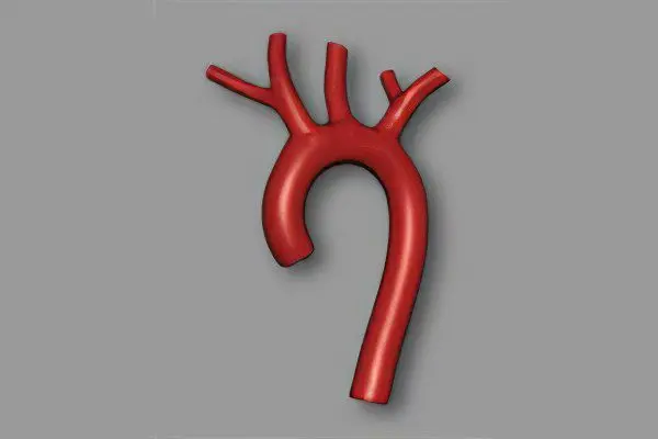
The largest artery in the human body is the aorta. This vessel of the systemic circulation exits the left ventricle and is subjected to high loads, since it receives the maximum pressure of blood flow. Therefore, an important and necessary property of the aorta is its elasticity and density.
Calcification is the deposition and gradual accumulation of calcium salts, forming calcified plaques, on the walls of the aortic valve. The danger of this process lies in the fact that as calcium penetrates into the walls of the vessel, they become more fragile, brittle and less elastic.
This, in turn, poses a threat to human life, because with an increase in blood pressure, the calcified walls of the vessel may not withstand and burst, which will lead to death. With calcification, calcium dissolved in body fluids (including blood) is deposited in tissues and blood vessels. This happens when there is too much calcium in the blood.
Causes of aortic calcification
The causes of an excessive amount of calcium salts in the blood, which significantly increase the likelihood of aortic calcification, may be the following:
With age, calcium is intensively washed out of the bones, getting into the blood in excess.
Various kidney diseases, in which the kidneys are not able to remove the required amount of calcium from the body and it enters the bloodstream.
In some pathological conditions of the intestine, there is also an increased absorption of calcium into the blood.
Sometimes there are deviations in the process of calcium absorption by bone tissue (complete absence or insufficient absorption). The remaining calcium enters the blood.
Also, the possible reasons for the development of aortic calcification include bad habits, malnutrition, stress, heart disease, heredity, physical inactivity, diabetes, obesity.
For the prevention and early diagnosis of aortic calcification, blood tests should be taken regularly. You can get a referral from a general practitioner or cardiologist. If the analysis shows an increased content of calcium in the blood, then you should look for the cause of this deviation.
aortic stenosis
Aortic stenosis is the fusion of the leaflets of the aortic valve. Since this pathology leads to a narrowing of the valve opening, the ejection of blood flow into the aorta is difficult, resulting in a significant increase in the load on the left ventricle, which leads to myocardial hypertrophy.
There are three forms of aortic stenosis: subvalvular, valvular, and supravalvular. Among patients with heart defects, aortic stenosis occurs in 84% of cases, and in men three times more often. Of all the forms of aortic stenosis, valvular is the most common. Also, aortic stenosis is congenital and acquired (90-97%). The supravalvular form is a congenital pathology.
Causes of aortic stenosis
The causes of acquired valvular or subvalvular stenosis can be aortic atherosclerosis, rheumatic valve disease, aortic calcification, and infective endocarditis. Sometimes the cause is diabetes mellitus, chronic kidney disease, Paget’s disease, systemic lupus erythematosus, or carcinoid syndrome.
Symptoms of aortic stenosis may not manifest themselves for a long time, especially at an early age. However, over time, the patient complains of squeezing, aching pain in the chest (in the heart), shortness of breath, fainting, palpitations, nausea, and fatigue. Since cardiac output is reduced, this can lead to loss of consciousness. In advanced stages, aortic stenosis can cause sudden onset of suffocation (cardiac asthma).
Also, in patients with aortic stenosis, the likelihood of sudden death increases, especially during physical exertion. Hypertrophied myocardium causes acute coronary insufficiency and arrhythmias that threaten the life of the patient.
Diagnosis of aortic stenosis
If symptoms of aortic stenosis appear, you should contact a cardiologist or start an examination with a therapist. With the help of a stethoscope, the doctor will listen to the noise above the aorta when the left ventricle contracts. An electrocardiogram reveals ventricular hypertrophy. An enlarged ventricle can also be detected with an x-ray. An echocardiogram uses ultrasound to assess the condition of the valves and the heart muscle.
Also, an informational research method is cardiac catheterization (insertion of a catheter into an artery). Gives information about intracardiac pressure, localization of narrowing, oxygen concentration in the blood, valve condition and the presence of a defect.
The most informative diagnostic method for making a decision on surgical intervention is an angiocardiographic study. It takes place in two stages: ventriculography and oortography.


