Contents
brain cyst – This is a common and rather dangerous disease that needs timely detection and high-quality treatment.
A cyst is a fluid-filled sac that can be located anywhere in the brain.
Most often, such cavities are formed in the arachnoid “network” that covers the cortex of the hemispheres, since its delicate layers are the most vulnerable to various inflammations and injuries.
This disease can be asymptomatic or cause pain and an unpleasant feeling of pressure in the patient.
In the case of an accurate diagnosis, the patient must follow all the recommendations of the doctor, and if necessary, agree to a surgical operation.
Signs and symptoms of a brain cyst

As a rule, a cyst can have a wide variety of sizes. Small formations usually do not manifest themselves in any way, and larger ones can put pressure on the membranes of the brain, as a result of which the patient has certain symptoms:
impaired vision or hearing;
headaches that are not amenable to stopping drugs;
insomnia;
violations of coordination;
partial paralysis of the limbs;
mental disorders;
muscle hypotonicity or hypertonicity;
noise in ears;
loss of consciousness and convulsions;
violations of the sensitivity of the skin;
pulsation in the head;
nausea and vomiting that does not bring relief;
limping;
feeling of pressure in the brain;
involuntary movements of the limbs;
pulsation of the fontanel and vomiting in infants.
It should be borne in mind that the clinical picture largely depends on where the formation is localized, since each part of the brain controls certain functions of the body. In addition, the occurrence of symptoms is significantly affected by the fact that exactly which area of uXNUMXbuXNUMXbthe brain the cyst exerts pressure on. For example, a mass in the cerebellum can cause problems with balance, change in gait, gestures, and even handwriting, and its appearance in areas responsible for motor or swallowing functions will cause difficulties in these areas. In addition, a cyst may not manifest itself in any way for a long time, and be found only in the process of conducting a tomographic study.
If the patient does not have the above signs of the disease, and the size of the cyst does not change in any way, then its presence may not affect his normal life at all, and it will be enough for him to confine himself to regular medical examinations. However, if the formation begins to increase, then this may be an indicator that the disease is progressing and the patient needs treatment.
Causes of a brain cyst

To begin with, consider how a cyst appears in the brain. In the space between the parietal and temporal lobes there is a fluid that, after a person has been injured, suffered a complex disease or surgery, can collect near the stuck together layers of the brain membrane, thus replacing the dead areas. If too much fluid accumulates, it can put pressure on these membranes, resulting in the formation of a cyst, and the patient has headaches.
Let us consider in more detail what causes can cause the appearance of this disease:
congenital disorders that are associated with an anomaly of intrauterine development of the fetus;
brain contusions, hematomas and fractures;
parasitic infections;
encephalitis;
meningitis;
degenerative and dystrophic transformations, as a result of which the brain tissue is replaced with cystic tissue;
violation of normal blood circulation in the brain.
If the underlying cause of the cyst is not identified, it may continue to grow in size. Its changes may be related to the following factors:
ongoing inflammation of the meninges;
fluid pressure on the dead part of the brain;
consequences of a concussion;
circulatory disturbance;
the emergence of new areas of damage after a stroke;
infectious disease, consequences of neuroinfection, encephalomyelitis, autoimmune process and multiple sclerosis.
Consequences, what is the danger of a brain cyst?
If an accurate diagnosis is not made to the patient in time and the correct treatment is not prescribed, this can lead to adverse consequences. Let’s consider how dangerous such a disease can be:
impaired coordination, as well as motor function;
hearing and vision problems;
hydrocephalus, manifested by excessive accumulation of cerebrospinal fluid in the ventricles of the brain;
encephalitis;
death.
As a rule, small formations that do not cause pain are detected during the diagnosis of other diseases and are cured with the help of medications without any complications. Large cysts that have an adverse effect on the structures of the brain that are located next to them usually need to be removed surgically.
Patients who have been diagnosed with this disease should not only treat it, but also observe certain preventive measures: do not overcool; beware of viral infections that can lead to complications; avoid situations that cause sudden changes in blood pressure, as well as give up bad habits such as alcohol abuse and smoking.
Types of brain cysts

This disease is classified into several types, each of which has its own characteristics and is characterized by certain symptoms. In modern medicine, the appearance of a cyst is not considered a pathology, but rather just an anomaly, in most cases not posing a threat to life. However, this mainly applies to congenital formations that are asymptomatic.
Primary cysts usually appear due to a violation of intrauterine development of the fetus or after the death of brain tissue due to intrapartum asphyxia. Acquired formations develop after inflammation, bleeding or bruising. In addition, they can be localized between the parts of the brain, or in its thickness in areas of dead tissues.
Arachnoid brain cyst located on its surface, between the layers of shells. Such a cavity filled with cerebrospinal fluid can be either congenital or occur under the influence of various factors. It is most common in male children and adolescents, and is much less common in women. As a rule, various inflammations and injuries lead to its appearance. If the pressure inside this formation becomes higher than intracranial pressure, then the cyst begins to compress the cerebral cortex.
An enlarged arachnoid cyst may be accompanied by symptoms such as nausea, vomiting, seizures, and hallucinations. It may become larger due to the fact that fluid pressure increases in it or because the patient continues to have inflammation of the meninges. If such a disease occurs, the patient must definitely consult a doctor, since rupture of the cyst can lead to death.
retrocerebellar brain cyst is a cavity filled with fluid, which is localized in its affected area. Unlike arachnoid formation, it does not occur outside, but in the thickness of the brain as a result of the death of gray matter cells. In order to prevent further damage to the brain, it is necessary to determine for what reason the cells died. A stroke can provoke the appearance of this formation; surgical operations on the brain; insufficiency of cerebral circulation; injury or inflammation, such as encephalitis. It should be borne in mind that new foci of infections and microstrokes can also cause cyst growth. In addition, it can increase due to the fact that circulatory disorders continue in the brain, and there is also a focus of infections that has a destructive effect.
Subarachnoid cyst of the brain usually detected by MRI. As a rule, such formations are congenital and are discovered by chance, in the process of carrying out diagnostic procedures. In order to assess its clinical significance, it is necessary to carefully check the patient for the presence of certain symptoms. This illness can be expressed by such signs as convulsions; feeling of unsteadiness or throbbing inside the skull.
If a retrocerebellar cyst of the brain begins to progress and grow, and is also accompanied by unpleasant symptoms, then in this case, a surgical operation may be required.
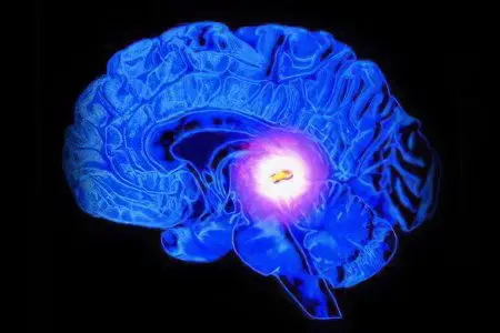
Pineal cyst of the brain – This is a cavity with a liquid that is formed at the junction of the hemispheres, in the pineal gland, which directly affects the endocrine system. The main reasons for its appearance can be factors such as echinococcosis or blockage of the excretory duct, leading to a violation of the outflow of melatonin.
Pineal brain cyst, which occurs in the epiphysis, is considered a fairly rare disease, it can lead to impaired metabolic processes, vision and coordination of movements. In addition, it often causes the development of hydrocephalus and encephalitis.
Cyst of the epiphysis of the brain It presents with symptoms such as headache, confusion, drowsiness, double vision, and difficulty walking. If the patient does not have the above symptoms, then there is a possibility that such a formation will not increase. This disease is found in the pineal gland in about four percent of people who undergo a CT scan for completely different reasons.
As a rule, at the first stage of this disease, doctors use medical methods of treatment, and constantly monitor the dynamics of its development, and if the disease is advanced, the formation is removed surgically. In the presence of pronounced symptoms, the patient should definitely consult a doctor in order to avoid various complications, such as dropsy, which can develop as a result of fluid accumulation.
Cerebral vascular plexus cyst It is in most cases a benign formation that appears at a certain stage of intrauterine development of the fetus. As a rule, such a cyst resolves on its own and is not a pathology. However, sometimes it can appear in newborns as a result of complications during pregnancy and childbirth or infection of the fetus. In some cases, such formation can lead to pathologies of other body systems.
In order to identify the presence of a cyst in infants, doctors perform a procedure such as neurosonography, which is completely harmless to the child. In adults, this disease is usually diagnosed with an ultrasound examination.
A subependymal cyst can occur in infants as a result of a violation of the blood circulation of the brain, as well as a lack of oxygen supply to it. This disease is considered more serious, and requires constant monitoring by doctors.
Liquor brain cyst – This is a formation that occurs between the sticky meninges. Its appearance is usually associated with inflammatory processes; stroke, meningitis, trauma or surgery. As a rule, this disease can be well diagnosed only in adulthood, since at an early stage of development the cyst is not well expressed, so it is difficult to identify it. Typical symptoms include nausea and vomiting; lack of coordination; mental disorders; convulsions, as well as partial paralysis of the limbs.
Lacunar cysts of the brain usually formed in the pons, in the subcortical nodes, and in more rare cases in the cerebellum and in the visual tubercles separated by white matter. There is an opinion that they appear as a result of atherosclerosis or age-related changes.
Porencephalic cyst of the brain occurs in the thickness of its tissues as a result of past infections. This disease can lead to very serious consequences, such as schizencephaly or hydrocephalus.
The colloidal cyst appears during the intrauterine development of the fetus and is of congenital origin. There is also a version that it is hereditary. Its main feature is that it blocks the outflow of fluid from the brain. This disease can occur without any symptoms throughout a person’s life, or be accompanied by signs such as headaches; epileptic seizures; high intracranial pressure or weakness in the legs. Symptoms of this disease usually appear in adulthood. It should be borne in mind that in some cases, a cyst can provoke the development of diseases such as cerebral hernia, hydrocephalus, and also cause death.
A dermoid cyst usually forms in the first weeks of fetal development inside the womb. Its cavity contains various elements of the ectoderm, sebaceous glands and hair follicles. Such a formation can increase quite quickly, so it is recommended to remove it surgically in order to avoid adverse consequences.
Treatment of a brain cyst

As a rule, cyst treatment is prescribed only after a complete diagnostic examination, which is performed using computed or magnetic resonance imaging, which allows you to see the clear contours of the formations, determine their size, as well as the degree of impact on the surrounding tissues.
It should be borne in mind that the presence of such cavities is not necessarily associated with cancer and usually responds well to treatment. During magnetic resonance imaging, a special contrast agent is injected into the patient, which makes it possible to determine what exactly is in his brain: a cyst or a malignant tumor. MRI is recommended to be performed repeatedly in order to constantly monitor the dynamics of the disease.
In order to prevent the patient from increasing cysts and the occurrence of new formations, it is necessary to identify the cause of their appearance. To this end, experts prescribe various studies, thanks to which you can find out what provoked the appearance of a cyst: infections, autoimmune diseases, or circulatory disorders. Let us consider in more detail the most common diagnostic methods:
Doppler study. This procedure is carried out in order to determine whether the vessels supplying arterial blood to the brain are narrowed. Violation of the blood supply can lead to the appearance of foci of death of the medulla, resulting in cysts.
Heart examination, ECG. This diagnostic method is carried out in order to detect heart failure.
Blood test for cholesterol and clotting. As a rule, high cholesterol and high clotting cause blockage of blood vessels, which, in turn, can lead to a disease such as a brain cyst.
Checking blood pressure. Its monitoring is carried out using a small device, on which the doctor records the patient’s pressure during the day on a memory card, and then all the information is read by a computer. If the patient has rises in pressure, then there is a possibility that this can cause a stroke and the appearance of post-stroke formations.
Blood test for infectious and autoimmune diseases. This examination is carried out in cases where there is a suspicion of arachnoiditis, neuroinfections or multiple sclerosis.
Methods for treating a brain cyst are selected based on the reasons that resulted in it. Emergency help is usually needed in the following cases:
recurring seizures;
hydrocephalus;
rapid increase in the size of the cyst;
hemorrhage;
damage to brain structures located next to the cyst.
As a rule, non-dynamic cysts of the brain do not require intervention, while dynamic ones are treated with the help of medicinal and surgical methods.
Traditional treatment involves the use of various medications, the main purpose of which is to eliminate the causes of the disease. Doctors may give patients medications that dissolve adhesions, such as caripain or longidase. In order to restore blood circulation, they prescribe drugs aimed at lowering the concentration of cholesterol, normalizing blood pressure and blood clotting.
It is possible to provide brain cells with the necessary amount of oxygen and glucose with the help of nootropics, for example, such as picamilon, pantogam, instenon. Antioxidants will help make cells more resistant to intracranial pressure. In addition, immunomodulatory, antibacterial and antiviral agents are sometimes used, which become necessary in case of detection of autoimmune and infectious diseases.
The appearance of arachnoiditis signals, first of all, that the patient’s immunity is greatly weakened, which is why it is necessary to actively engage in the restoration of protective forces. In order to select a consistent and safe course of immunomodulatory and anti-infective treatment, you need to take a blood test. As a rule, all medications are prescribed in courses lasting about three months, repeated twice a year.
Removal of a brain cyst
Radical treatment of a brain cyst involves its removal by surgery. For this purpose, the following methods are used:
Shunting. This method of treatment is carried out using a drainage tube. Through the device, the cavity is emptied, as a result of which its walls begin to subside and “overgrow”. However, it should be borne in mind that this method increases the likelihood of infection, especially if the shunt is in the skull for a long time.
Endoscopy. Such operations aimed at removing the cyst with punctures usually pass without complications. They are associated with a small proportion of injuries, but they also have certain contraindications, for example, they are not recommended for patients with impaired vision. In addition, this method is not used for every type of cyst.
Trepanation of the skull. This operation is considered quite effective, but it must be borne in mind that during its implementation the risk of brain injury is very high.
For the treatment of newborn babies in the departments of pediatric neurosurgery, similar operations are performed, but only if the cyst progresses and increases, as a result of which there is a danger to the development and life of the child. During the surgical operation, computer monitoring is carried out, which allows doctors to monitor its progress and quickly make the right decisions.
Surgery can avoid many of the adverse effects that a brain cyst can cause, such as mental disorders, developmental delays, headaches, and loss of speech, vision, or hearing. If after the operation the patient does not have any complications, his hospitalization is about four days, and after discharge from the hospital, he must undergo regular examinations with his attending physician.
Timely treatment of this disease in most cases can prevent its recurrence and reduce the risk of various complications, especially if you go to a clinic that uses modern medical equipment, as well as professional and qualified specialists.


