


Scientists from the USA developed and implemented a new type of endoscopic camera, guided by a weak magnetic field and transmitting the image wirelessly. It will significantly facilitate the diagnosis of the gastrointestinal tract in hospitals, informed the websites of Eurekalert and BWH.
Scientists from Brigham and Women’s Hospital (BWH) in Boston have successfully completed the first series of clinical trials of the new endoscopic camera, constructed according to the principle of “device on the pill”. This camera, inspired by constructions from science-fiction movies, has the ability to “swim in the human body” and gives clinicians an insight into previously difficult to reach parts of the body and internal organs.
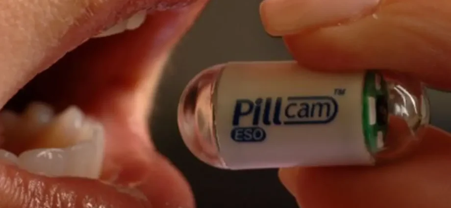
The new device differs from the currently used “camera in a nutshell”. Currently, an automatic camera is used, which moves along with the food or fluid in the digestive tract and takes pictures at a planned time interval. Access to them can only be gained after the expulsion and disassembly of the camera. In addition, it is a multi-purpose device only.
The new camera in a nutshell is smaller than the one used so far. It has an integrated chip including a high-resolution camera and a wireless communication system, ensuring transmission distance up to 2 m. The control is carried out by an MRI device for magnetic resonance, generating a magnetic field, acting on a pill equipped with small magnetic domains. Hence, the capsule can be “held” by an organ or its passage through the gastrointestinal tract can be accelerated so that it reaches the place of interest to doctors faster. The image can be transmitted via a wireless module in real time.
As stated in the description of the research by Noby Hat, a scientist from the Radiology Department at BWH, who led the research and trials of the new endoscopic capsule, the aim of the project was to construct such a device that would enable real-time image transmission, enabling clinicians to make a correct diagnosis in just one analytical procedure without risk to the patient.
The BWH team performed the first tests in a tank full of water and in a tube system partially filled with water, simulating the human digestive system. The next stage of the tests will be to test the camera during hospital diagnostics, which will probably take place in February this year.
Scientists would similarly use nanomaterials and control the magnetic field generated by MRI to create a system for the safe transport of drugs directly to cancerous tumors or damage to internal organs.