Contents
Bassin
The pelvis (from Latin pelvis) is a bony belt which supports the weight of the body and which forms the junction between the trunk and the lower limbs.
Anatomy of the pelvis
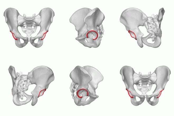
The pelvis, or pelvis, is a belt of bone located below the abdomen that supports the spine. It is made from the association of the two coxal bones (hip bone or iliac bone), the sacrum and the coccyx. The hip bones are themselves the result of the fusion of three bones: ilium, ischium and pubis.
The hip bones join behind the sacrum, by the wings of the ilium, at the level of the sacroiliac joints. The upper edge of the wing is the iliac crest, it is the point of insertion of the abdominal muscles. The iliac spines are palpable when you put your hands on your hips.
The two hip bones meet at the front at the level of the pubis. They join together by the pubic symphysis. In a seated position, we are posed on the ischio-pubic branches (branch of the pubis and ischium).
The pelvis is attached with the lower limbs at the level of the hip or coxofemoral joint: the acetabulum (or acetabulum), a C-shaped joint cavity, receives the head of the femur.
A funnel-shaped cavity, the pelvis is divided into two regions: the large pelvis and the small pelvis. The large basin is the upper part, delimited by the wings of the ilium. The small basin is located under these wings.
The cavity is delimited by two openings:
- the upper strait which is the upper opening of the basin. It marks the transition between the large and the small pelvis. It fits into the space delimited from front to back by the upper edge of the pubic symphysis, the arched lines and the promontory of the sacrum (upper edge) (3).
- The lower strait is the lower opening of the basin. It forms a diamond. It is limited anteriorly by the inferior border of the pubic symphysis, on the sides by the ischiopubic branches and ischial tuberosities, and finally posteriorly by the tip of the coccyx (4).
In pregnant women, the dimensions of the basin and the straits are important data to anticipate the passage of the baby. The sacroiliac joints and the pubic symphysis also gain a little flexibility through the action of hormones to promote childbirth.
There are differences between the male and female pools. The female pelvis is:
- Wider and more rounded,
- Shallower,
- Its pubic arch is more rounded because the angle formed is greater,
- The sacrum is shorter and the coccyx straighter.
The pelvis is the place of insertion of various muscles: the muscles of the abdominal wall, those of the buttocks, the lower back and most of the muscles of the thighs.
The pelvis is an area that is heavily irrigated by numerous vessels: the internal iliac artery which is divided in particular into the rectal, pudendal or ilio-lumbar artery. The pelvic veins include among others the internal and external iliac vein, common, rectal …
The pelvic cavity is richly innervated by: the lumbar plexus (eg: femoral nerve, lateral skin of the thigh), the sacral plexus (eg: posterior skin nerve of the thigh, sciatica), the pudendal plexus (eg: pudendal nerve, penis , clitoris) and the coccygeal plexus (eg: sacral, coccygeal, genitofemoral nerve). These nerves are intended for the viscera of the cavity (genitals, rectum, anus, etc.) and the muscles of the abdomen, pelvic and upper limbs (thigh).
Pelvic physiology
The main role of the pelvis is to support the weight of the upper body. It also protects the internal genitals, bladder and part of the large intestine. The hip bones also articulate with the thigh bone, the femur, which allows walking.
Pelvic pathologies and pain
Fracture of the pelvis : it can affect the bone at any level but three areas are generally the most at risk: the sacrum, the pubic symphysis or the acetabulum (the head of the femur sinks into the pelvis and breaks it). The fracture is either caused by a violent shock (road accident, etc.) or a fall coupled with bone fragility (eg osteoporosis) in elderly subjects. The viscera, vessels, nerves and muscles of the pelvis can be affected during a fracture and cause sequelae (nervous, urinary, etc.).
Hip pain : they have various origins. However, in people over 50, they are most often linked to osteoarthritis. Often, pain associated with a hip disorder will be “misleading”, localized for example in the groin, buttock, or even in the leg or knee. Conversely, the pain can be felt in the hip and actually come from a more distant point (the back or the groin, in particular).
Pudendal neuralgia : affection of the pudendal nerve which innervates the region of the pelvis (urinary tract, anus, rectum, genitals…). It is characterized by chronic pain (burning sensation, numbness) aggravated by sitting. It generally affects people between 50 and 70 years old and the cause of this pathology is not clearly identified: it can be a compression of the nerve or its enclavement in different areas (pinched between two ligaments, in the canal under pubic …) or by a tumor for example. Neuralgia can also be caused by excessive use of the bicycle or childbirth.
Pelvic movements during childbirth
Specific movements in the sacroiliac joints that allow vaginal delivery:
- Counter-nutation movement: a verticalization of the sacrum (retreat and elevation of the promontory) occurs when it is associated with an advancement and a lowering of the coccyx and a separation of the iliac wings. These movements have the effect of enlarging the upper strait * and reducing the lower strait **.
- Nutation movement: the reverse movement occurs: advancement and lowering of the promontory of the sacrum, retreat and elevation of the coccyx and approximation of the iliac wings. These movements have the consequence of enlarging the lower strait and narrowing the upper strait.
Hip osteoarthritis (or coxarthrosis) : corresponds to the wear of the cartilage at the level of the joint between the head of the femur and the hip bone. This progressive destruction of the cartilage is manifested by pain in the joint. There are no treatments that would allow cartilage regrowth. Hip osteoarthritis, or coxarthrosis, affects about 3% of adults.
Treatments and prevention of the pelvis
The elderly represent a population at risk of pelvic fractures because they are more exposed to falls and their bones are more fragile. The same is true for people with osteoporosis.
Preventing a fall is not easy, but it is advisable to consume foods rich in calcium and vitamin D to strengthen bones and fight against osteoporosis. For older people, it is important to eliminate any obstacle in their environment that could be the cause of a violent fall (removal of the mats) and to adapt their behavior (installation of bars in the toilets, wearing of shoes that hold the foot). It is also advisable to avoid the practice of sports at risk of violent falls (parachuting, horse riding, etc.) (10).
Pelvic examinations
Clinical examination: if a pelvic fracture is suspected, the doctor will first carry out a clinical examination. For example, he will check whether there is pain when mobilizing the sacroiliac joints (between the ilium and the sacrum) or a deformity of a lower limb.
Radiography: medical imaging technique that uses X-rays. Frontal and lateral radiography makes it possible to visualize the bone structures and organs contained in the pelvis and to highlight a fracture for example.
MRI (magnetic resonance imaging): medical examination for diagnostic purposes carried out using a large cylindrical device in which a magnetic field and radio waves are produced. Where radiography does not allow it, it reproduces very precise images. It is particularly used in cases of hip and pubic pain. To visualize the organs, MRI can be combined with the injection of contrast product.
Pelvic ultrasound: imaging technique that relies on the use of ultrasound to visualize the internal structure of an organ. In the case of the pelvis, ultrasound makes it possible to visualize the organs of the cavity (bladder, ovary, prostate, vessels, etc.). In women, it is a common examination for the follow-up of pregnancy.
Scanner: diagnostic imaging technique which consists of “scanning” a given region of the body in order to create cross-sectional images, thanks to the use of an X-ray beam. The term “scanner” is actually name of the medical device, but it is commonly used to name the exam. We also speak of computed tomography or computed tomography. In the case of the pelvis, a CT scan can be used to look for a fracture not visible on an x-ray or for a pelvimetric measurement (pelvic dimensions) in pregnant women.
History and symbolism of the basin
For a long time, having a large pelvis was associated with fertility and as such was considered a criterion of seduction.
Nowadays, on the contrary, a narrow pelvis is preferred to the image of the famous size 36.


