Contents

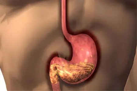
Atrophic gastroduodenitis can become a consequence of chronic inflammation of the mucous membrane of the stomach and duodenum. A characteristic feature of this disease is the destruction of the secretory glands that produce gastric juice. Instead of juice, the degenerated glands produce mucus. This form of gastroduodenitis is considered a precancerous condition and develops against a background of low acidity. Approximately 12% of all cases of atrophic gastroduodenitis are associated with the introduction of the bacterium Helicobacter pylori into the patient’s body.
After infection of the body with the bacterium Helicobacter, the concentration of gastric juice, which protects the gastrointestinal tract from dangerous infections, changes. The pathological process from the stomach very quickly passes into the duodenum, which disrupts the process of digestion of the food bolus. During progressive inflammation of the gastric mucosa, secretory, or parietal, glands are lost, and metaplasia of individual sections occurs.
If the atrophic process occupies at least 20% of the entire area of the stomach, it can be said with absolute certainty that there is cancer. According to medical research, every eighth case of atrophic gastroduodenitis ends with oncological pathology, while in other forms of gastritis this probability is 5 times lower.
With timely diagnosis of the disease, after 5 years of high-quality treatment, the areas of metaplasia are significantly reduced, and the mucous membrane of the stomach and duodenum is restored.
Causes of atrophic gastroduodenitis

There are two main causes leading to atrophy of the mucous membrane of the stomach and duodenum:
An autoimmune process in which the G cells of the secretory glands are damaged by their own immune antibodies;
Prolonged stay in the gastrointestinal tract bacteria Helicobacter pylori.
During the autoimmune process, antibodies are mistaken for foreign tissues by cells of their own secretory glands. The acidity of the gastric juice gradually decreases, the parietal glands begin to produce mucus instead of hydrochloric acid. These processes lead to the impossibility of absorption of iron and vitamins by the walls of the stomach and duodenum, the development of anemia. Attachment of the bacterium Helicobacter accelerates the formation of sites of metaplasia.
The introduction of an infection leads to cell damage, due to which free radicals penetrate into them. The glands change their structure, their cells become precancerous. This is how intestinal metaplasia develops, when sections of the gastric mucosa acquire the properties of the small intestine and large intestine epithelium. These transformations increase the likelihood of gastric adenocarcinoma.
Factors contributing to the development of the disease:
Physical and mental overstrain;
Hereditary predisposition to diseases of the gastrointestinal tract;
Side effects of drugs;
stress;
Alcohol abuse;
Occupational diseases;
Somatic diseases in a chronic form.
Symptoms of atrophic gastroduodenitis

The disease develops slowly, begins from the bottom of the stomach, gradually moving to other parts of the mucous membrane. Vivid symptoms of the disease may not manifest themselves at first, which creates obstacles for early diagnosis and timely treatment.
Signs of an anemic syndrome that occurs due to malabsorption of vitamins and iron:
Weakness;
Fast fatiguability;
Drowsiness;
Pale skin and mucous membranes;
Burning and pain in the tongue;
Lacquered surface of the tongue;
dry hair;
Brittle nails;
Stitching pains in the heart;
Shortness of breath under any load.
Signs of dyspeptic syndrome associated with indigestion:
Heaviness in the stomach;
Aching pain in the projection of the epigastrium;
Heartburn;
Burping;
Nausea;
Vomiting of recently eaten food, mucus and bile;
Decreased appetite;
Alternating constipation and diarrhea;
Bad breath and bad taste in the mouth in the morning;
Gray coating on tongue, imprint of teeth on it.
Violation of digestion leads to a sharp decrease in body weight, in advanced cases – to dystrophy. Obstacles to the absorption of vitamins cause a decrease in immunity, frequent colds and infectious diseases.
Diagnostics

The most informative modern method for determining the form and stage of gastroduodenitis is the hematological diagnostic panel. This study helps to establish the degree of damage to the secretory glands and the level of metaplasia of the mucosal epithelium.
Parameters determined by the study:
Gastrin level-17;
The level of pepsinogen-1 and pepsinogen-2;
Histamine level-17;
The ratio of indicators.
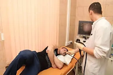
Another informative study is EGDES with methylene blue staining of the mucosa to assess the area of epithelial metaplasia. During an endoscopic examination, a tissue biopsy of all altered sections of the mucous membrane is taken.
Additional methods for diagnosing atrophic gastritis:
Gastrography;
Intragastric ph-metry;
Daily measurement of acidity;
MSCT (multispiral computed tomography) – if gastric cancer is suspected;
Determination of the presence or absence of the bacterium Helicobacter pylori (breath test, ELISA test, PCR reaction).
Treatment of atrophic gastroduodenitis

The goal of the treatment of atrophic gastroduodenitis is to prevent the further development of intestinal metaplasia, the destruction of the epithelium and its transformation into atypical cells (cancerous transformations). This goal can be achieved within 5 years of careful therapy.
A prerequisite for a full-fledged treatment is dietary nutrition. Food should be sparing in composition, temperature, mechanical structure. After a short period of time, it is allowed to include low-concentrated lemon, cranberry, cabbage juice in the diet. Bananas are the only acceptable fruits for this diet. Food should not be either cold or hot, the diet should be frequent meals, small portions. It is absolutely unacceptable during treatment and after it smoking, drinking alcohol in any dose.
Drugs for the treatment of atrophic gastroduodenitis:
Antibiotics to eradicate the bacterium Helicobacter pylori;
proton pump inhibitors;
The preparation is bismuth;
Glucocorticosteroids;
Iron preparations;
Vitamins;
Enzymes;
Mineral waters with a high content of minerals;
Gastroprotectors;
Antacids;
Stimulators of cellular regeneration;
Means that stimulate peristalsis.
In addition, physiotherapy treatment (electrophoresis, magnetotherapy, thermal procedures), sanatorium treatment at a balneological resort are used.
Prophylaxis and prognosis
To prevent atrophic gastroduodenitis, acute and chronic diseases of the gastrointestinal tract should be treated in a timely manner, and the principles of rational nutrition should be followed. Early access to a gastroenterologist and quality treatment will help prevent complications.
For elderly patients, the prognosis for the development of the disease is worse than for younger patients. At the age of over 50 years, mucosal atrophy most often ends with malignancy. If one course of treatment does not relieve the disease, it should be repeated.


