Contents

X-ray therapy for heel spurs is a method of treating a disease by exposing problem areas to x-rays.
X-rays are widely used in medicine not only for the diagnosis of various pathologies, but also for their therapy. The technology is electromagnetic radiation with a short wavelength and high energy. The rays have the ability to penetrate deeply into tissues and produce a therapeutic effect.
Most often, X-ray therapy is used to rid a person of oncological diseases. Doctors using this type of radiation affect benign tumors, remove warts and other skin formations.
When undergoing X-ray therapy, it is always worth remembering that X-rays are radiation and, in a certain concentration, can harm a person. At the same time, such therapy should not be abandoned if it is recommended by a doctor. After all, X-rays are harmful to health only with a prolonged and intense effect on the body. X-ray therapy is suitable for the treatment of heel spurs, since the affected area is in an easily accessible place and can be targeted.
The essence of the method
When exposed to x-rays on the heel spur, the body is not affected by harmful rays. That is why X-ray therapy is a very popular method of treating this disease, especially in the case when the question of surgical intervention arises. It is recommended to use it only when conservative treatment has not given the desired effect.
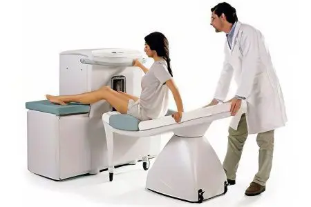
For the procedure of heel spur radiotherapy, special equipment is used, which is a source of x-rays. The doctor directs him to the place where the bone growth is located.
As a result of the influence of X-rays, the nerve endings are blocked and the pain recedes. It is known that it is pain that is the leading symptom in the presence of a heel spur, and it is they that reduce the quality of human life. Low doses of radiation can eliminate this pain. Naturally, the spur itself will remain in place, but it will no longer bother a person.
A session of exposure to x-rays on the heel spur lasts about 10 minutes. In order to completely block the nerve endings, it is necessary to undergo 10 procedures. A full course of treatment is a guarantee that the pain will recede forever.
The procedure is performed after the patient has been examined by an orthopedic doctor and after passing all the necessary examinations (X-ray, MRI, UAC). During the session, the patient is placed on the couch, and the leg is raised. A special tube is placed on the heel area, which is the source of x-rays. After the session, experts recommend unloading the sore leg. To do this, you need to wear shoes with arch supports, get rid of excess and weight, use canes or crutches, and perform other actions aimed at facilitating the functioning of the foot.
Therapeutic effects that are achieved through exposure to x-rays:
analgesic effect. It will be noticeable after the first procedure, it is achieved by blocking sensitive receptors;
Anti-inflammatory effect. It is achieved due to the normalization of blood circulation in the affected area. As a result, metabolic processes are normalized, the synthesis of prostaglandins responsible for the inflammatory reaction is reduced;
Hypoallergenic effect. It is achieved due to the fact that tissues become less sensitive to the effects of potential allergens;
regeneration effect. It is achieved due to the destruction of old cells and their accelerated recovery.
Benefits of heel spur radiotherapy
The procedure is absolutely painless and comfortable for the patient.
One session is short in time and takes no more than 10 minutes.
To eliminate pain, it is enough to complete one course of 10 procedures.
In addition to relieving pain, X-ray therapy can reduce inflammation and eliminate allergic reactions.
Treatment does not require the patient to stay in the hospital, immediately after the procedure, he can continue to do his own thing.
The action of X-rays is distinguished by strict purposefulness.
The procedure avoids surgery and possible complications from it.
X-ray therapy is often the last treatment before the patient is sent to the operating table. The effectiveness of the irradiation will be affected by the severity of the pathology, the age of the patient and the duration of the course.
It is possible to repeat the course if necessary, but this can be done no earlier than after 3 months. As a rule, this is enough to consolidate the result and save the patient from pain for a long time.
Disadvantages and contraindications of heel spur radiotherapy
X-ray therapy for heel spurs is contraindicated for pregnant women. The fact is that x-rays can adversely affect the condition of the fetus.
Despite the fact that the majority of patients indicate a positive effect of undergoing radiotherapy, no studies have been conducted in this regard. Just as there was no purposeful long-term monitoring of patients who underwent such treatment. Therefore, it is not possible to speak unambiguously about the absolute safety of the method.
In this regard, any specialist begins therapy for heel spurs with simpler methods of treatment. In the event that they do not help, or there are contraindications for their implementation, they resort to X-ray therapy.


