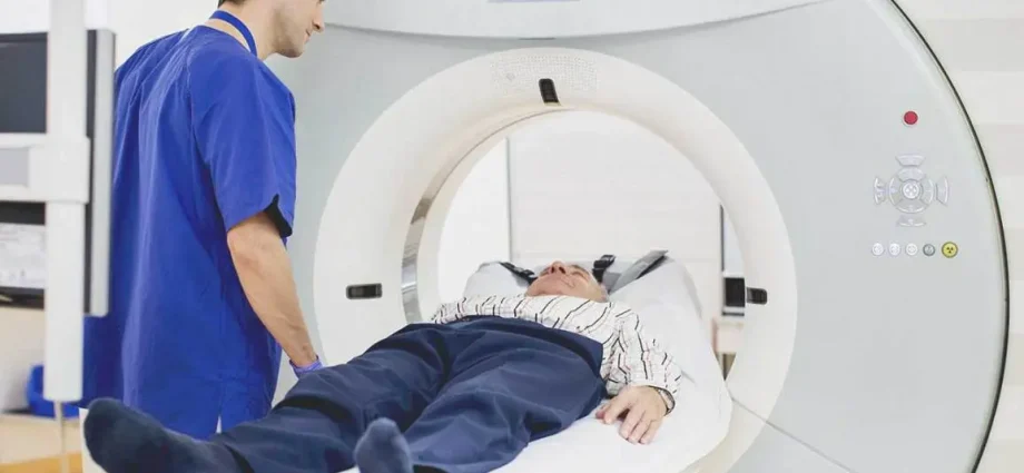
Lung cancer does not show symptoms until an advanced stage. However, there is a way to detect it at an early stage, when the changes will not be revealed by an X-ray image. It’s a low-dose computed tomography. There is a program in Poland that offers it to former and current smokers. What can they gain from such research, says Prof. Ewa Sierko from the Department of Oncology of the Medical University of Białystok.
- Lung cancer is one of the most common and poor prognosis malignancies. In 95 percent. is the result of smoking a cigarette
- Low-dose computed tomography (NDTK) makes it possible to detect lung cancer early.
- Currently, recruitment to this preventive program is ongoing throughout Poland. It will run until the end of June
- We are asking everyone not to postpone their decision and report now – appeals oncologist-radiotherapist prof. Ewa Sierko
- The program is aimed at, inter alia, to patients who are or have been smokers and have quit smoking less than 15 years ago
- The test is completely safe for the body. What is low-dose computed tomography?
- More information can be found on the Onet homepage.
She graduated from the Medical Academy in Białystok. She is a specialist in oncological radiotherapy. Currently, he heads the Radiotherapy Department of the First Oncology Center in Białystok and is a professor at the Oncology Clinic of the Medical University in Białystok.
He looks after the low-dose computed tomography (NDTK) program in the Lubelskie and Podlaskie voivodships. She is a co-author of over 262 scientific papers, one monograph and 46 chapters in Polish and English medical textbooks.
Beata Igielska / Zdrowie.pap: The low-dose computed tomography (NDTK) program was ready before the pandemic, but its outbreak suspended recruitment for several months. How is it now?
Prof. From Sierko: In Podlaskie Voivodeship, the program was supposed to start in April, but when the pandemic broke out, recruitment stopped for several months. At the moment, for a change, we have a very strong interest in the program. However, there are still vacancies and recruitment will only last until the end of June this year.
Nationally, only 13 percent are missing. willing to take part in the program, so you will probably be able to recruit the number of people you need?
I am convinced that we will be able to recruit the missing number of patients. We had 200 vacancies two weeks ago. We are currently strongly promoting the program. It is worth remembering that low-dose computed tomography makes it possible to detect lung cancer early. For patients who are diagnosed with lung cancer at an early stage, it gives a significant improvement in treatment outcomes, which translates into a longer life. Therefore, we are asking everyone who is willing not to postpone their decisions and report now, by contacting medical institutions that support the program in the country. The Bialystok Cancer Center represents the eastern area and supervises the Podlaskie and Lubelskie voivodeships. All of Poland takes part in this program.
The rest of the text is below the video.
Please tell me what it is?
The program, which is designed to detect lung cancer at an early clinical stage, is aimed at patients who are or were smokers and quit smoking less than 15 years ago. People who smoke for at least 20 pack years are eligible for the program, i.e. those who have smoked one pack of cigarettes a day for 20 years, or two packs a day for 10 years, or half a pack of cigarettes a day for 40 years.
Where did such criteria come from?
Hence, 95 percent. lung cancer is the result of smoking. Screening programs, and this one includes them, are always aimed at a group of people who is particularly exposed to the development of a given cancer. Therefore, if someone has smoked for 20 pack years and is 55-74 years old, they can participate in a low-dose chest computed tomography program.
Another criterion is that they must be patients who do not have symptoms suggestive of lung cancer, such as dyspnoea, hemoptysis, cough, repeated recurrent pneumonia, or suffering from this cancer. Those who have previously been diagnosed with lung cancer are already under the control of pulmonary or oncology centers.
Why is low-dose computed tomography recognized in the world as a screening method for the early detection of lung cancer?
Because it actually detects this cancer at an early stage in which the cancer is not yet showing any symptoms. In a certain group of patients, participation in the program is possible five years earlier, i.e. from the age of 50. This applies to people working with silica, nickel, asbestos and diesel exhaust fumes (e.g. as car mechanics). But it also applies to people living in the vicinity of coal smoke or working as chimney sweeps with soot, or those whose family history has had lung cancer in first-degree relatives, i.e. parents or siblings. It also includes patients with chronic obstructive pulmonary disease, or those who have developed tobacco-dependent cancer in a different location, e.g. head and neck cancer or bladder cancer.
Buy a kit of diagnostic tests:
- oncology package for women
- oncology package for men
What can you see in low-emission tomography?
We even see a tiny lump in the lung. In the past, we only had an X-ray. It showed us much larger lung tumors that were no longer eligible for surgery. Large randomized trials conducted in the United States on over 50 of patients showed that in the risk group among patients who smoked more than 30 years a pack of cigarettes a day, the detection of a nodule at an earlier stage of cancer development reduces the risk of death by 20%. This is a lot, because currently most lung cancers are diagnosed in the advanced stage of the disease, when it is no longer possible to undergo surgery or stereotaxic radiotherapy. In the later stages of the disease, the treatment results are much worse.
Is Low Dose Chest Computed Tomography Safe for the Patient?
It is completely safe for the body, because the dose of X-rays during the examination is small. It lasts a few minutes, the intravenous contrast is not administered, which additionally makes the examination safe and completely painless.
What else, apart from the size of the lung nodules, can be assessed by such a tomography?
You can also assess the condition of the coronary vessels, see if there is emphysema. If we detect problems other than a lung nodule, the patient receives information from us and can start diagnosing other diseases. This short study gives you the chance to improve your health and extend your life.
I would like to emphasize that the early stage of lung cancer, when surgery can be performed or stereotaxic radiotherapy can be used, gives a chance of five-year survival (in oncology it is assumed that five-year survival means curing the cancer – ed.) In over 70% of patients. patients. This is a very good effect. Each subsequent stage of the disease development worsens the chances of a complete recovery. When there are metastases to the bones, liver, brain – not more than a few percent of patients survive five years.
We are very happy with this program, especially after the last two years, when, due to the pandemic, the pulmonary departments were occupied by covid patients, which delayed the diagnosis of lung cancer. Moreover, it was difficult to get to a pulmonologist. In a pandemic, 15 percent was issued. fewer DiLO cards (Fast Oncology Pathway). And in the case of lung cancer, delay in diagnosis by three months means a decrease in cure rate by 10%, delay in diagnosis by six months means a decrease in cure rate by as much as 30%. During the pandemic, the diagnosis rate of advanced lung cancer increased by 20-30%.
These numbers are very disturbing …
Exactly. Therefore, the low-dose chest computed tomography program “has fallen from the sky” because it offers an opportunity for early cancer detection. Thanks to it, we achieve similar results as we have now achieved in breast cancer. Women with a large tumor that can be felt through the skin used to seek treatment for breast cancer, but now most breast cancers are found on screening mammography.
Low-dose computed tomography translates into tangible results – prolonged survival. In the United States, this program is standard, with us it is a pilot program, but we hope that it will be available to everyone, like mammography, financed by the National Health Fund.
So this program is about shifting the treatment of lung cancer patients from stage three and four to stage one and two?
More specifically, it is about moving the diagnosis of the disease to the first stage of clinical advancement, when we achieve the best treatment results. The cancer most often shows symptoms when it is in its third or fourth stage. No treatment at this late stage can replace early cancer detection and offer such a high chance of prolonging survival and a good quality of life for the patient.
The tomography shows even tiny lumps that most likely won’t even turn into cancer. In their opinion, do you use a specific algorithm, e.g. artificial intelligence?
Yes. Radiologists use an algorithm. Patients in the program have at least one tomography performed. If we immediately notice that the nodule is malignant, the patient is referred for diagnostics. However, depending on specific indicators and guidelines, the patient has a tomography performed in three months or in six months. We observe whether the nodule changes into a nodule with evident malignancy during this time. People who do not have a lump on their first CT scan will have a repeat examination in a year.
After the publication of the results of the study, Nelson and radiologists working in the low-dose computed tomography program base their diagnosis on the spatial size of the nodule and calculate its volume. The perception of lung nodules as spatial structures is better translated into the assessment of whether a lump is malignant, as opposed to a two-dimensional assessment. Computed tomography scans the lungs in layers every few millimeters. Until now, often only two nodule dimensions have been reported on a single transverse lung scan.
Today, is every CT scanner capable of detecting a malignant nodule with a low-emission X-ray dose?
Yes, any CT scanner can do low dose tomography, it’s even easier. Low-dose CT examinations have been used for many years, e.g. in dentistry to plan implantological procedures or in hematology to assess whether there are no bone metastases. To use this method to detect lung cancer, however, we needed evidence from research. These were performed in the United States and confirmed that such a study is effective in diagnostics and translates into a reduction in mortality in the population.
And can the scanner window be set so that you can see, for example, the outline of a kidney?
We also see the outline of the upper part of the kidney and adrenal gland, because the lower lungs are concave and some abdominal structures are visible on CT scans.
And if something in this disturbing window appears, then after this tomography you can send patients to other specialists for further diagnostics?
If we see a breast tumor on tomography, we send it to a breast disease treatment clinic, if we see problems with the coronary vessels, the patient may be referred to a cardiologist. We refer to a pulmonologist when a patient has bronchiectasis or emphysema. The patient may be referred to a specialist directly or to a family doctor with information about the result of the tomography.
As I understand it, a whole team of specialists is necessary for the smooth functioning of the program, almost like in the case of DiLO: radiologists, thoracic surgeons, oncologists, pulmonologists?
This program works before the DiLO card is inserted, i.e. before making a decision about the need for oncological treatment. Radiologists working with it are properly trained. Result visits are performed by pulmonologists and thoracic surgeons. We oncologists work closely with them.
Detailed information about the program on the website lungcheck
Strong menstrual pain is not always “so beautiful” or a woman’s hypersensitivity. Endometriosis may be behind such a symptom. What is this disease and how is living with it? Listen to the podcast about endometriosis by Patrycja Furs – Endo-girl.
Beata Igielska, zdrowie.pap.pl


